Scientific Harrovian

UNCOVERING THE SCIENTIFIC WORLD
WILL OUR FUTURE SURGEONS BE ROBOTS?
HOW TO REVERSE HEARING LOSS? WHO WANTS TO LOOK YOUNG?
MAXIMIZING PHOTOSYNTHESIS
Issue viii
2022-2023
Harrow International School Hong Kong
“ “
About the
Scientific Harrovian 2023
The Scientific Harrovian is the student run Science Department magazine, which provides a platform for students to showcase their research and writing talents, and for more experienced pupils to guide authors and to develop skills to help them prepare for life in higher education and beyond.
All images, unless otherwise specified, are obtained from Unsplash or Pexels Portrait photos credited to Dora Gan of Photography Society
3 Prologue
Ms. McCrohan
a message from Head of Biology
I would like to thank, and congratulate, all of the writers, editors and illustrators on the completion of this outstanding edition. You are each important cogs in the Scientific Harrovian machine, without each of you this edition simply would not have happened. The team has been run passionately by two exceptional students, Judy Sheng and Kevin Wu, who have motivated the team to produce well researched and written pieces across a range of subjects. The final piece was put together through the tireless efforts of Cyrus Tsui, our Chief Design Officer, resulting in an edition that is visually stunning. I hope everyone who picks up a copy of this Scientific Harrovian enjoys it too.

4 Scientific Harrovian 2022

a message from
Judy Sheng
Editor-in-chief
I am delighted to welcome you to issue VIII-i of the Scientific Harrovian!
This year’s theme is ‘Uncovering the Scientific world’, with articles covering from spider silk to quantum computing. Each article is bound to give you a glimpse into unique realms of science, and I hope you enjoy them as much as I did!
Our team has made a great start to the year with countless hours of work put in by our writers, illustrators, editors, and members of the executive team. My greatest thanks go to all our contributors for their commitment and effort. It has been a huge pleasure to seeing the team grow and watching this publication come together as old and new members join us on another enthralling expedition into the Scientific World.
A special thanks to the members of our executive team, especially Cyrus Tsui (Chief Design Officer) and Grace Zhu (Deputy Design Officer) for putting together this amazing piece of work. And of course, to Kevin Wu, our Deputy Editor-in-Chief who led the team and helped coordinate this issue. None of this could have come together without each of their hard work.
I hope you enjoy!
a message from
Kevin Wu
Deputy Editor-in-chief
WELCOME!
As our life gradually recovers from the pandemic and returns to normality as a result of the vaccine, it is crucial for us to realise how the applications of sciences shape our world today. Hence, the theme of Edition VIII-i of the Scientific Harrovian is Uncovering the Scientific World, focusing on the contemporary applications of science in our day-to-day life and its potential in the future.

With this issue being the time bearing such a great responsibility as the Head Editor in chief, I cannot express my gratitude enough towards Judy for not only trusting me but also for guiding me to make this issue possible. I would also like to thank Cyrus, the Chief design officer, for his expertise in designing and I’m truly amazed at how he managed to put everything together from scratch. Lastly, I would like to thank all the head editors, editors, and writers for dedicating your already precious time to attending weekly lunchtime meetings and contributing your part to the issue. This issue really wouldn’t be possible without any of you!
Enjoy!
5 Prologue
the TEAM


Editor-in-Chief
Deputy Editor-in-Chief
Chief Design Officer
Deputy Chief Design Officer
Biology Head Editor
Chemistry Head Editor
Physics Head Editors


Judy Sheng
Year 13 Gellhorn
Kevin Wu
Year 12 Sun
Cyrus Tsui
Year 12 Peel
Grace Zhu
Year 11 Gellhorn
Jenny Park
Year 12 Wu
Adrian Lau
Year 12 Peel
Sky Lee
Year 12 Churchill
Emma Chua
Year 12 Gellhorn
Technology Head Editor
Emma Chua
Year 12 Gellhorn


6 Scientific Harrovian 2022
writers
Jasmine Wong
Year 13 Keller
Sen Yi Mok
Year 11 Shaftsbury
Gloria Kan
Year 12 Anderson
Kate Xiao
Year 12 Gellhorn
Daniel Kan
Year 11 Shaftsbury
Audrey Lai
Year 12 Gellhorn
editors
Jack Wei
Year 6 Banks
Tracy Zhang
Year 9 Wu
Davyn Kwok
Year 7 Darwin
Peony Sham
Year 12 Anderson
Cyrus Tsui
Year 12 Peel
Ashlee Kwan
Year 11 Wu
Edward Wei
Year 13 Peel
Clarence Chen
Year 12 Sun
Emma Chua
Year 12 Gellhorn
illustrators
Tracy Zhang
Year 9 Wu
Cindy Min
Year 11 Gellhorn
Cyrus Tsui
Year 12 Peel
Bernice Ho
Year 9 Anderson
Sky Lee
Year 12 Shaftsbury
Gloria Siu
Year 12 Keller
Fabiola Chong
Year 12 Keller
Bess Chau
Year 12 Gellhorn
Elaine Zhang
Year 12 Gellhorn
Karen Li
Year 12 Gellhorn
Carol Yeung
Year 13 Keller
Bernice Ho
Year 9 Anderson
Callum Sanders
Year 12 Shaftsbury
Rachel Pabaru
Year 12 Wu
Andrew Hung
Year 11 Churchill
Eileen Wu
Year 8 Nightingale
Sky Lee
Year 12 Shaftsbury
Zhaoping Sun
Year 10 Churchill
Grace Zhu
Year 11 Gellhorn
Ethan Lan
Year 9 Churchill
Aiden Lan
Year 6 Shackleton
Lara McWilliam
Year 13 Keller
Ivy Sham
Year 9 Anderson
Andrea Lee
Year 11 Gellhorn
Valerie Ho
Year 11 Anderson
Janus Guo
Year 6 Banks
Adrian Lau
Year 12 Peel
Jenny Park
Year 12 Wu
Daniel Kan
Year 11 Shaftesbury
Bernard Ho
Year 7 Banks
Branda Mak
Year 12 Wu
Toby Li
Year 12 Peel
Katy Shiu
Year 9 Wu
Charlize Mui
Year 10 Wu
Tisha Handa
Year 11 Anderson
Toby Li
Year 12 Peel
Jenny Yin
Year 12 Wu
Marco Lee
Year 10 Churchill
Jolie Lai
Year 12 Wu
Pockmen Deng
Year 11 Shaftesbury
Lorina Lee
Year 9 Anderson
Branda Mak
Year 12 Wu
Callum Sanders
Year 12 Shaftsbury
Sky Lee
Year 12 Shaftsbury
7 Prologue

8 Scientific Harrovian 2022

CONTENTS Physics and Technology Chemistry and Biology Will Our Future Surgeons be Robots? ---------------- 12 Alphafold ------------------------------------------------ 20 The Fundamentals of Quantum Computing ---------- 27 The Speed of Light and its Significance --------------- 35 Starlink Satellite Probability Collision Simulation - 43 Who Wants to Look Young? 46 New Treatments for Cancer? 51 How can sustainability be achieved through the arts of building design? 55 Medicinal Applications of Spider’s silk --------------- 61 The Power of Stem Cells -------------------------------- 69 Photosynthesis ------------------------------------------ 90 Synthesised Meat ---------------------------------------- 97 Maximimising photosynthesis ---------------------- 103 The technology that surgical robots need to improve and the current solution 111 How to reverse hearing loss? 115 Introduction to CRISPR 119 Spanish flu vs Covid-19 pandemic ------------------- 125 Social Prescribing ------------------------------------- 132 9 Prologue


10 Scientific Harrovian 2022

PHYSICS and TECHNOLOGY
11 Physics and Technology
WILL OUR FUTURE SURGEONS BE ROBOTS?
By Daniel Kan

12 Scientific Harrovian 2022
1. Introduction
Robotic surgery is the use of mechanical arms carrying surgical instruments that are controlled by a surgeon. Robotic surgery is generally used with minimally invasive surgeries, which use small incisions instead of the traditional open procedures.
2. The History of Robotic Surgery
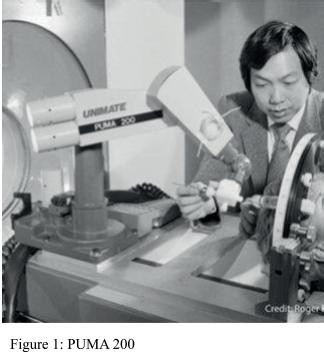
The first application of robots in surgery was in 1985, when a Programmable Universal Machine for Assembly (PUMA 200) was used to perform a neurosurgical biopsy. [1] It was further adapted by The Robotics Center at Imperial College into the PROBOT [2], which was specifically designed to perform a transurethral resection of the prostate (TURP), a procedure that involves cutting away a section of the prostate. The PROBOT allows a surgeon to specify a volume of the prostate, which would automatically be cut by a rotating blade [3].
In 1992, the ROBODOC system was developed and became the first active robot system to achieve a formal FDA approval. This was used to improve the precision of hip replacement surgery. The ROBODOC system consists of a preoperative surgical planning workstation called ORTHODOC and a five-axis robotic arm to carry out the plan [4].
During the next decade, the field of robotic surgery underwent a paradigm shift in which research was more focused on the “master-slave” concept, where a surgeon would remotely control the movements of a robot from a distant workstation.
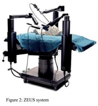
In 1989, a company called Computer Motion created a robotic platform called Automated Endoscopic System for Optimal Position (AESOP). This consisted of a robotic arm that held an endoscope which removed the need for an assistant to hold it. This had multiple benefits, such as not fatiginge during long procedures (unlike if an assistant was holding it), more stability, and less personnel required to be present in the operation.
Initially, the AESOP 1000 (approved in 1994) was controlled by pedals, but later the AESOP 2000 could be controlled using a voice control system. The final platform, the AESOP HR, also had voice control of other functions such as operating room lighting and movement of the operating table [5].
In 1998, the AESOP system was modified and relaunched as the ZEUS operating system, which had arms and surgical instruments that could
13 Physics and Technology
be remotely controlled by the surgeon. The ZEUS operating system had three arms: one was an AESOP camera system that was controlled by voice, the other two arms held surgical instruments that could be controlled using handles. In 2001, the first transatlantic surgery was carried out using the ZEUS system, where a surgeon from New York performed a cholecystectomy (removal of gallbladder) on a patient in France [6].
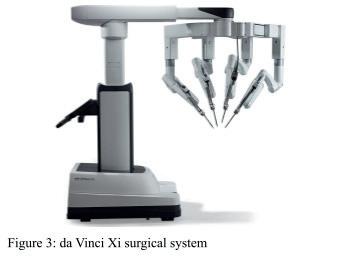
At the same time that the ZEUS system was being developed, another company called Intuitive Surgical was developing their own surgical robot. Their first prototype was called Lenny and had three arms: two for holding instruments and one for the camera. The second generation of robots was Mona, which was the first robotic surgical system to be used in human trials. However, in 1998, Intuitive surgical developed the da Vinci system, which would later become the most successful robotic surgery platform that is still used to this day. The first da Vinci robot had three arms: one that held the camera and the rest would hold instruments. These arms could rotate with seven degrees of freedom and two degrees of axial rotation – a significant selling point compared to other systems. The two companies Computer Motion and Intuitive Surgical then merged in 2003, discontinued the ZEUS system and worked together to improve the da Vinci system [7]. In 2000, the da Vinci system gained FDA approval for clinical use, and 2 years later a version with 4 arms was also approved. In 2006, the da Vinci S platform was released, with a 3D HD camera and an interactive touch screen display. In 2009, the da Vinci Si platform was released with dual console surgery, allowing two surgeons to operate at once. This optimised each surgeon’s potential as well as introduced a way to train non-expert surgeons. The Si system also had other improvements such as a better image system and real time fluorescence imaging. In 2014, the da Vinci Xi platform was created, as well as the da Vinci SP system, which had a single port and only required one incision.
3. How Does Robotic Surgery Work?
3.1. Robotic Surgery vs Minimally Invasive Surgery vs Open Surgery
Traditionally, open surgery requires the surgeon to make a large incision using a scalpel to view the necessary organs. Minimally invasive surgery (MIS) uses several small incisions and a laparoscope, which has a small camera attached to it, to allow the surgeon to examine the organs. MIS is generally less painful and has a faster recovery period compared to open surgery [8]. Robotic Surgery or Robot Assisted Surgery is generally associated with MIS and uses robotic arms that are controlled by the surgeon. The robotic arms hold a camera and surgical instruments. Robotic surgery has multiple advantages such as a greater range of motion and dexterity(ability to delicately manipulate with hands and fingers) for the surgeon [9]. It usually also has a faster recovery time.
3.2. How current Robotic Surgery works - The da Vinci system
The da Vinci system has 3 components: the surgeon console, the patient cart and the vision cart. These components follow the “master-slave” concept, where the surgeon console is the “master interface” and the patient cart holds the “slave manipulators” that hold the surgical instruments. The vision cart makes communication between components possible and supports the 3D HD vision system [10].
14 Scientific Harrovian 2022
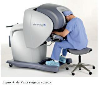
The surgeon console allows the surgeon to see inside the patient and control the manipulators. The stereo viewer gives the surgeon a 3D-HD view which immerses the surgeon in the surgical field, something that was lost when doing traditional minimally invasive surgery. The two master controllers allow the surgeon to control the instruments and endoscope. The surgeon can use their hands to move the master controllers, and the actions will be replicated by the manipulators. The manipulators are designed to allow a natural range of motion, dexterity and ergonomic comfort (when using and holding) [11]. Through these controllers, the surgeon’s hand tremors can be filtered out from the electronic signal or scaled down. The surgeon console also has left side and right side pods, which contain controls such as ergonomic controls, the power button, and an emergency stop button [12]. The surgeon console also has a footswitch panel which allows the surgeon to control different things using their feet without having to remove their head from the 3D viewer.
The Patient cart is the operative component of the system and has four arms that hold all the instruments and endoscope. During the operation, instruments and endoscopes are swapped by the assistant surgeon.
The Vision cart holds electronic equipment for visualisation. It includes a light source to illuminate the surgical site, soft ware processing units to process the video images and send it to the 3D viewer and touchscreen.
3.3. Visualisation
The da Vinci Surgical system uses endoscopes to allow the surgeon to visualise the area they are operating on. These endoscopes transmit white-light to form images that only show the visible surfaces of the organs [13]. Recently, there have been further innovations with other techniques such as the Firefly Fluorescence imaging. This works by injecting a fluorescent agent into the bloodstream which will emit light when excited. This is then excited using a corresponding excitation light source and the fluorescence can be detected using a specific detector. The most widely used fluorescent agent is indocyanine green (ICG), which rapidly binds to plasma proteins in the blood. When the ICG fluoresces, the image detected can be combined with the white light image to allow the surgeon to see vasculature and tissue perfusion. One way that ICG is removed from the blood is by secreting it into bile at the liver. This can allow the surgeon to visualise the structures of the bile duct.

15 Physics and Technology
There are many other techniques that can be used for visualisation. Dynamic view expansion or mosaicing can offer a wider field of view [14]. Narrow Band Imaging uses specific filters to modify white light images to increase contrast which allows surgeons to more clearly view a certain part of the tissue [15]. Tomographic imaging can be used before or during surgery which uses penetrating waves to provide cross sectional images beyond the surface tissue.
3.4. The Surgical Instruments
The surgical instruments used by the da Vinci system have an articulated wrist mechanism called EndoWrist which allows it to have more dexterity and a greater range of motion [10]. EndoWrist instruments include endoscopic dissectors, scissors, scalpels, forceps, needle holders, needle drivers, retractors, bipolar and monopolar energy instruments, suction irrigation instruments, staplers, and more [16].

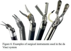
The staplers are used in transection and resection by placing multiple rows of staples then transecting the tissue with a knife blade, cleanly cutting the tissue without any bleeding [13]. The stapler is controlled by the foot pedals at the surgeon console.
There are also instruments that use energy. These are split into monopolar and bipolar instruments. Monopolar is when the current passes from the electrode to the target tissue then to a return pad and back to the generator to complete the circuit. Bipolar is when the current passes from one side of the instrument to the other side of the instrument, and only the tissue between the instrument is affected.
There are three monopolar instruments used in the da Vinci system: the hook, the scissors, and the spatula [16]. The most common monopolar instrument is the hook, which allows the surgeon to dissect and apply energy to a certain area. The scissors allow the surgeon to precisely dissect tissue in restricted spaces. The spatula is used for desiccation (drying out of cells) over a wide area [17].
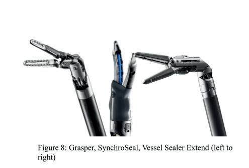
Several bipolar instruments are used in the da Vinci system. The bipolar grasper is used to grasp and retract tissue, and can also be used for hemostasis of small blood vessels. The bipolar forceps can also be used for hemostasis of small vessels, as well as being used to cut, although this is rarely used [17]. The Vessel sealer extend and Vessel sealer can seal and cut vessels. They do this by precisely applying pressure and energy to control the temperature, causing soft tissue proteins to denature and melting the inside walls of the vessel together. Once the vessel is sealed, a mechanical knife can be used to cut through the vessel. The vessel sealer can also be used for dissection, which decreases operative time by removing the need to change instruments. SynchroSeal is another instrument that can also seal vessels. It is more efficient than vessel sealer since it only requires a single pedal press to seal and cut as opposed to two pedal presses. However, Vessel sealer extend can seal and cut vessels up to 7 mm in diameter, whilst SynchroSeal can only seal and cut vessels up to 5mm in diameter.
16 Scientific Harrovian 2022
4. Current Appliations of Robotic Surgery
Currently, robot assisted surgery is used across a wide range of surgical specialties. Whilst robotic surgery is most commonly performed in urological, gynaecological and gastrointestinal surgery [18], robotic surgery has also seen use in many other specialties.
4.1. Urological surgery
Due to the depth of the pelvis and small size of the structures [19], prostatectomy was one of the first surgical operations to widely adopt robotic surgery. This allows surgeons to more easily guide the instruments to the required location (eg. prostate, kidneys), which is more advantageous when compared to open surgery or traditional laparoscopic techniques. As well as prostatectomy, robotic surgery has also been used in nephrectomies and adrenalectomies. Overall, robotic surgery has been widely adopted for urologic surgery, especially for performing prostatectomies.
4.2. Gynaecological surgery
Robotic surgery has been used to perform many operations in gynaecology. It is estimated that over 60% of hysterectomy procedures (removal of the uterus) were done robotically [21]. Robotic surgery has also been used in myomectomy (removal of uterine fibroids whilst preserving the uterus), tubal reanastomosis, and pelvic and paraaortic lymph node dissection [21].
4.3. Application in gastrointestinal surgery
The increased quality of the images produced by the endoscope and increased precision of its instrument is important in the treatment of gastrointestinal cancer [22]. This includes removal of cancer in organs such as the stomach, liver, gallbladder, small bladder, adrenal, colon, and others [19].
4.4. Other surgical fields
Robotic surgery has also seen use in other surgical fields. In otolaryngological (head and neck) surgery, robotic surgery allows for smaller incisions whilst still allowing the surgeon to have clear visualisation and dexterity. In neurosurgery, robotic surgery allows surgeons to surpass the limits of human dexterity on a microscopic scale, although it does have its limitations such as speed and lack of sense of touch. In cardiothoracic surgery, robotic surgery has been used for mitral valve surgery, repairing atrial septal defect, anastomosis on an arrested heart, anastomosis on a beating heart, and more [19].
5. Future Applications of Robotic Surgery
5.1. Telerobotic Surgery
Telerobotic surgery, also known as remote surgery, is robotic surgery where the surgeon is in a distant place and communicates through a wireless network. This was initially explored by NASA who wanted a type of surgery that could be performed in space, but it can also be applied on earth so patients do not have to travel long distances [23]. Telerobotic surgery allows surgeons from around the world to perform surgery in rural areas or places with surgeon shortage. It can even allow for collaboration of multiple surgeons which can be used to enhance care, as well as for training [24].
However, latency time (delay) has been a significant drawback, as too much time delay can lead to inaccuracies. Developments in 5G technology can be useful for reducing this. There can also be problems with cyber security, cost, and legal issues across country borders [24].
17 Physics and Technology
Despite some of these issues, in 2019 researchers in China successfully performed 12 telerobotic spinal surgeries using 5G, all of which were successful. In all of these, the master surgeon was in a different province to the patient. The researchers concluded that using 5G telerobotic surgery was “accurate, safe, and reliable” [25].
5.2. Nanorobotic surgery
Nanorobots are tiny robots that can move around the patient’s entire body through the bloodstream (including capillaries) to access different cells. This can be used for highly precise surgery down to the cellular level, as well as accessing hard to reach places. Nanorobots can take many forms, such as nanodrillers, micro-grippers, micro bullets, and more [26]. Aside from surgery, nanorobots can also be used for targeted drug delivery, diagnosis, detection, biopsies, imaging, 3D printing, and more [27]. However, nanorobotics is just beginning and there still are many challenges that need to be overcome.
5.3. Autonomous robots
In the future, it is possible that robots will be able to perform surgeries autonomously without the control of a human surgeon. To do this, the robot will have to be able to interpret visual and physical data, then decide what to do and carry it out [28]. It will also need to be able to adapt to different situations in real time. To achieve this, various machine learning algorithms will need to be used for receiving and interpreting data, as well as being “taught” how to actually perform the surgery.
Although current robotic surgeries are still done by humans, recently, a robot successfully performed an intestinal anastomosis on a pig without any direct human assistance using the Smart Tissue Autonomous Robot (STAR) [29].
6. Future Applications of Robotic Surgery
In conclusion, robotic surgery is a type of minimally invasive surgery that uses robotic arms to perform surgery. Currently, it uses the “master-slave” concept where a surgeon directly controls robotic manipulators that hold surgical instruments and an endoscope. There are many benefits of robotic surgery including more dexterity, better visualisation, and faster recovery times. However, robotic surgery is very expensive, which is why it hasn’t been as widely adopted as it could be. Fields where robotic surgery is used the most are: urological surgery, gynaecological surgery, and gastrointestinal surgery. In the future, it could be used for telerobotic surgery where the surgeon controls the robot remotely, nanorobotic surgery where nanorobots move through the bloodstream, or autonomous surgery where the robot performs without any human assistance.
18 Scientific Harrovian 2022
7. Bibiliography
[1] Kwoh, Y.S., et al. “A Robot with Improved Absolute Positioning Accuracy for CT Guided Stereotactic Brain Surgery.” IEEE Transactions on Biomedical Engineering, vol. 35, no. 2, 1988, pp. 153–160., https://doi.org/10.1109/10.1354.
[2] “Probot.” Imperial College London, www.imperial.ac.uk/mechatronics-in-medicine/research/probot/.
[3] “The Method of Cutting the Prostate with the Robot.” Imperial College London, www.imperial.ac.uk/mechatronics-in-medicine/research/probot/cutting/.
[4] Bargar, William L., et al. “Primary and Revision Total Hip Replacement Using the Robodoc?? System.” Clinical Orthopaedics and Related Research, vol. 354, 1998, pp. 82–91., doi:10.1097/00003086-199809000-00011.
[5] MORRELL, ANDRE LUIZ, et al. “The History of Robotic Surgery and Its Evolution: When Illusion Becomes Reality.” Revista Do Colégio Brasileiro De Cirurgiões, vol. 48, 2021, doi:10.1590/0100-6991e-20202798.
[6] Marescaux, Jacques, et al. “Transatlantic Robot-Assisted Telesurgery.” Nature, vol. 413, no. 6854, 2001, pp. 379–380., doi:10.1038/35096636.
[7] Lane, Tim. “A Short History of Robotic Surgery.” The Annals of The Royal College of Surgeons of England, vol. 100, no. 6_sup, 2018, pp. 5–7., doi:10.1308/rcsann.supp1.5.
[8] “Open Surgery vs Laparoscopic Surgery: Which Is the Best Procedure?” Far North Surgery, www.farnorthsurgery.com/blog/open-surgery-vs-laparoscopic-surgery.
[9] “What Is Robotic Surgery?” UCLA Health System, www.uclahealth.org/medical-services/robotic-surgery/what-robotic-surgery.
[10] “About Da Vinci Systems.” Da Vinci Surgery | Da Vinci Surgical System | Robotic Technology, www.davincisurgery.com/da-vinci-systems/about-davinci-systems.
[11] Mishra, R.K., System Components - World Laparoscopy Hospital. www.laparoscopyhospital.com/Book/Ch-03.pdf.
[12] Intuitive Surgical, da Vinci Si surgical system User Manual, Intuitive Surgical
[13] Azizian, Mahdi, et al. “The Da Vinci Surgical System.” The Encyclopedia of Medical Robotics, 2018, pp. 3–28., doi:10.1142/9789813232266_0001.
[14] Lerotic, Mirna, et al. “Dynamic View Expansion for Enhanced Navigation in Natural Orifice Transluminal Endoscopic Surgery.” Medical Image Computing and Computer-Assisted Intervention – MICCAI 2008, 2008, pp. 467–475., doi:10.1007/978-3-540-85990-1_56.
[15] Barbeiro, Sandra, et al. “Narrow-Band Imaging: Clinical Application in Gastrointestinal Endoscopy.” GE - Portuguese Journal of Gastroenterology, vol. 26, no. 1, 2018, pp. 40–53., doi:10.1159/000487470.
[16] Da Vinci X & Da Vinci XI Instrument & Accessory Catalogue - Intuitive.com. Intuitive Surgical, Mar. 2022, www.intuitive.com/en-gb/-/media/ISI/ Intuitive/Pdf/da-vinci-x-xi-instrument-accessory-catalogue-1075017.pdf.
[17] Wikiel, Krzysztof J., et al. “Energy in Robotic Surgery.” Annals of Laparoscopic and Endoscopic Surgery, vol. 6, 2021, pp. 9–9., doi:10.21037/ ales.2020.03.06.
[18] Anderson, Jamie E., et al. “The First National Examination of Outcomes and Trends in Robotic Surgery in the United States.” Journal of the American College of Surgeons, vol. 215, no. 1, July 2012, pp. 107–114., doi:10.1016/j.jamcollsurg.2012.02.005.
[19] Shah, Jay, et al. “The History of Robotics in Surgical Specialties.” American Journal of Robotic Surgery, vol. 1, no. 1, 2014, pp. 12–20., doi:10.1166/ ajrs.2014.1006.
[20] Bharathan, Rasiah, et al. “Operating Room of the Future.” Best Practice & Research Clinical Obstetrics & Gynaecology, vol. 27, no. 3, 21 Dec. 2012, pp. 311–322., doi:10.1016/j.bpobgyn.2012.11.003.
[21] Leddy, Laura, et al. “Robotic Surgery: Applications and Cost Effectiveness.” Open Access Surgery, 2 Sept. 2010, p. 99., doi:10.2147/oas.s10422.
[22] Ohuchida, Kenoki. “Robotic Surgery in Gastrointestinal Surgery.” Cyborg and Bionic Systems, vol. 2020, 2020, pp. 1–7., doi:10.34133/2020/9724807.
[23] Mohan, Anmol et al. “Telesurgery and Robotics: An Improved and Efficient Era.” Cureus vol. 13,3 e14124. 26 Mar. 2021, doi:10.7759/cureus.14124
[24] Choi, Paul J, et al. “Telesurgery: Past, Present, and Future.” Cureus, 2018, doi:10.7759/cureus.2716.
[25] Tian, Wei et al. “Telerobotic Spinal Surgery Based on 5G Network: The First 12 Cases.” Neurospine vol. 17,1 (2020): 114-120. doi:10.14245/ ns.1938454.227
[26] Li, Jinxing et al. “Micro/Nanorobots for Biomedicine: Delivery, Surgery, Sensing, and Detoxification.” Science robotics vol. 2,4 (2017): eaam6431. doi:10.1126/scirobotics.aam6431
[27] Eggleton, Benjamin. “Nanorobotic Surgery.” Nanorobotic Surgery, The University of Sydney, www.sydney.edu.au/nano/our-research/research-programs/nanorobotic-surgery.html.
[28] Panesar, Sandip, et al. “Artificial Intelligence and the Future of Surgical Robotics.” Annals of Surgery, vol. 270, no. 2, Aug. 2019, pp. 223–226., doi:10.1097/sla.0000000000003262.
[29] Saeidi, H., et al. “Autonomous Robotic Laparoscopic Surgery for Intestinal Anastomosis.” Science Robotics, vol. 7, no. 62, 26 Jan. 2022, doi:10.1126/ scirobotics.abj2908.
Figure 1: https://www.researchgate.net/figure/Puma-200-the-first-robot-used-for-assisting-human-neurosurgery-1985-12_fig2_290495998
Figure 2: https://www.researchgate.net/figure/ZEUS-robotic-system-first-robotic-system-to-combine-instrument-and-camera-control_fig3_51437277
Figure 3: http://www.rsalinas.com/davinci-si-1-1
Figure 4: https://www.advancedurologyinstitute.com/da-vinci-surgical-system/
Figure 5: https://www.ourmidland.com/news/article/Firefly-glow-improves-visibility-in-surgery-6946851.php
Figure 6: https://entokey.com/the-da-vinci-system-technology-and-surgical-analysis/ Figure 7 and 8: https://www.intuitive.com/
19 Physics and Technology

ALPHAFOLD
What Is It and Does it Really Solve the Protein Folding Problem?
By Gloria Kan
20 Scientific Harrovian 2022
1. Introduction
Proteins are everywhere, from specifically shaped enzymes that catalyse metabolic processes, to the fibrous, connective tissue made of collagen present in just about every organ in the body, to the body’s chemical messengers, hormones, that are secreted from exocrine glands and travel in the bloodstream. They play an irreplaceably crucial role in our daily lives, impacting our appearance, our actions, and most importantly, our survival!
Hence, AlphaFold is an extremely useful AI, as it can predict how chains of amino acids can fold into complex 3D structures, namely secondary, tertiary and quaternary, as protein functions are highly reliant on their shapes. However, before we go into the specifics of how AlphaFold works, we should understand why it is necessary first.
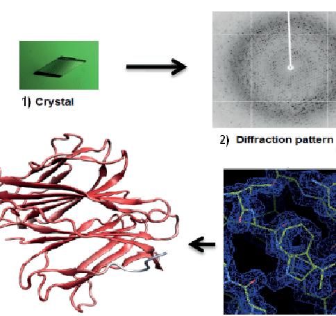
Traditionally, X-ray crystallography has always been the principal technique used to determine the complex 3D structure of proteins, especially for small, soluble proteins [6]. It goes through the following stages:
1. Crystallisation; this is when a pure, highly concentrated sample of protein is crystallised. pH, concentration, temperature and additive inclusion are controlled to optimise the yield and quality of protein crystals suitable to determine the structure of a protein.
2. A single X ray beam is generated by accelerating electrons caused by an electron striking a copper anode, and it is passed through slits that are approximately 0.1-0.3 mm wide, which causes diffraction (the spreading out of waves) [2].
3. A CCD (charge-coupled device) collects the X-ray diffraction images; it is generally preferred over conventional X-ray film or imaging plates due to its high level of sensitivity and the fact that the images can be collected rapidly (in a matter of seconds).
4. Resolution is calculated; it is important for it to be 1.5-3 Å (1Å is 1x10^−10 m), ensuring all amino acid side chains can be identified. (For reference, a carbon bond is approximately 1.5 Å) [2].
5. Data can then be collected for an electron density map, and analysed for a final structure.
(Figure 1: the structure of a potential plant disease resistance protein [14])
21 Physics and Technology
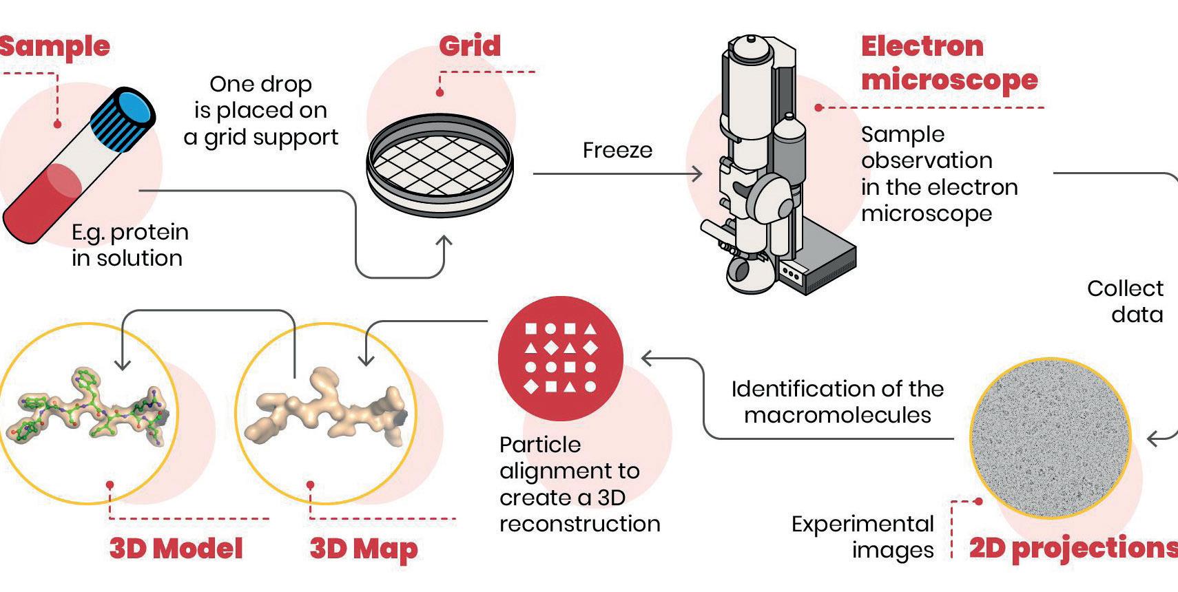
Another rapidly advancing procedure is single-particle cryo-electron microscopy (cryo-EM) [1], a method that is most prominent in identifying larger protein structures. Its procedures are [3]:
1. Apply a pure protein sample to a grid with small holes in its film.
2. Put the grid into a cryogen (a gas at a very low temperature; an example may be liquid nitrogen) to flash-freeze and trap particles in a thin film of vitreous ice. This is to protect the sample from any damage caused by radiation.
3. In the transmission electron microscope, a low electron dose is used to reduce damage done to the sample. As signals can be weak, many particles from the sample are analysed by a computer algorithm, to form one image of the particle; this is known as particle averaging.
4. Many 2D views of the protein obtained from different angles are processed to align images and merge data for a 3D map. Instead of having to convert electrons to photons, direct detectors can now detect electrons directly, allowing images to be recorded like a movie, resulting in a higher resolution (as motion correction can be used to reduce radiation drift) .
5. The main protein sequences are then fitted into a 3D map for a 3D model of the protein.
 (Figure 2: the stages of X-ray crystallography [2])
22 Scientific Harrovian 2022
(Figure 3: the process of cryo-electron microscopy [15])
There is also NMR (Nuclear Magnetic Resonance) spectroscopy [4]. Its method is as follows:
1. Put the sample into a strong magnetic field —the stronger it is, the more detailed molecules can be studied, causing some atomic nuclei to act as small magnets.
2. When a range of frequencies is applied to it, the nuclei resonate at specific frequencies.
3. These frequencies of the nuclei are measured and analysed on a spectrum where intensity increases with larger resonating nuclei.
4. The value of the frequency gives information about the relative positions of atoms.
5. By examining the cross peaks of intensity, scientists can determine the 3D structures of proteins.
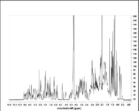
 (Figure 4: example of a one-dimensional NMR spectrum of rubredoxin, a small protein [16])
23 Physics and Technology
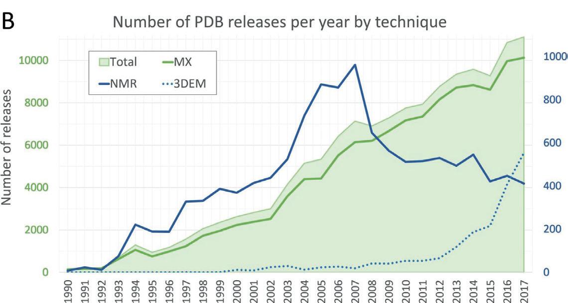
(Figure 5: NMR is nuclear magnetic resonance spectroscopy, 3DEM is 3D electron microscopy and Mx is macromolecular crystallography (essentially cryo-EM) [5])
Unfortunately, these methods can be very time-consuming and expensive, as it can take anytime from a few months to years of painstaking effort for one protein structure to be identified by a research lab. Before AlphaFold, it took over 50 years of arduous experimental efforts for scientists to identify the structures of approximately 100,000 proteins (around 50,000 being human protein structures). However, despite this impressive number, it is only about 17% of the human proteins, and many structures only cover a fragment of the sequence [8]. Thus, AlphaFold is a great advancement in the realm of biology, as it can predict protein structures quickly with a fairly high level of confidence.
2. The history of AlphaFold (A7D and CASP13)
It all started when DeepMind, the developer of AlphaFold (previously known as A7D), entered CASP13 (the critical assessment of protein structure prediction), a prestigious competition that has been running since 1994 [7]. For fairness, they use a system of blind testing, a method of testing the entrants’ modelling systems with the structures (of recently discovered proteins) that haven’t been input into the protein data bank yet.
In CASP13, DeepMind’s model structure, A7D, secured them the top place in the competition. They used three different free-modelling methods; a GDT-net, a gradient descent to predict backbone structure, and a distance potential fragment assembly, with the gradient descent method achieving the highest scores and high accuracy structures (GDT_TS, a measure of the similarity of a model and a predicted structure scores of 70 or higher out of a 100 being predicted in) being predicted for 11/43 GDT-net proteins [10].
Its overall design combined predictions of several neural networks that estimated the distances between the carbon atoms of pairs of residues, since residues may have been positioned closely together even if they weren’t close in the sequence of amino acids. As a result, a contact map with data about distances and angles for each residue could be used to predict a 3D structure.
However, with A7D, overfitting was present; interactions between residues were over accounted for, and, as a result, models were believed to have more secondary structures when it wasn’t necessarily true (i.e. the AI believed that the protein had more alpha helices and beta pleated sheets than interactions between tertiary or quaternary structures).
3. CASP14 and AlphaFold
Since CASP13, AlphaFold has gone through drastic improvements. First, the AlphaFold network can now directly predict the 3D coordinates of a given protein using the primary amino acid sequence as inputs [9]. It starts by employing Multiple Sequence Alignments (MSAs) with different regions weighted by importance (attention) through repeated layers of a novel neural network block (also known as Evoformer) [11]. Then, in the trunk, it extracts information about the relationships between the protein sequence and template structure, producing a Seq N x Res N array (where Seq N is the number of sequences and Res N is the number of residues); residues are also known as unique R groups in an amino acid giving it its properties. Furthermore, in the trunk, there are regular updates about the relationship between the sequence-residue and residue-residue edges of a graph in order to achieve consistency and fit the constraints.
In the head, the structure module treats the protein as if it is a residue gas moving around the network to generate the protein’s 3D structure. Initially, rotations are set to identity and all positions are at the origin, but a protein structure is swiftly developed. And, unlike A7D, end-to-end folding is used instead of gradient descent, and the 3D transformer directly operates on a rigid 3D backbone using pair representation and the original sequence row from the MSA to build the side chains.
After that, there is the refinement step, which ‘refines’, or improves the accuracy and stereochemical qualities of the protein, and a step known as relaxation. As the result isn’t guaranteed to obey all stereochemical 24 Scientific Harrovian 2022
restraints, violations of any constraints (especially peptide bond geometry, as it is less controlled in the structure module) can be resolved with coordinate restrained gradient descent.
As for the results achieved in CASP14, AlphaFold 2 received an overall z-score (indicating similarity between two protein models) of 244.0217, while the next best group scored 90.8241. Moreover, AlphaFold managed the best prediction out of all participants for 88 out of 97 of targets, with levels of accuracy equivalent to experimental x-ray crystallography.
4. Limitations
However, AlphaFold cannot perfectly predict protein structures. Some predictions made by AlphaFold fail to reach a high level of accuracy in CASP14. T1047s1-D1, for example, only managed a median accuracy value of 50.47 (out of 5 models) with a long beta sheet at a completely incorrect angle from the domain (the rest of the structure), and this is thought to be due to it having “a very high oligomerization state (quaternary structure)” and a “lack of other intra-domain structure” [12]. Thus, it can be discerned that it is very difficult for AlphaFold to predict proteins consisting of one or more polypeptides.
Furthermore, AlphaFold can only predict backbone and side chain structure for a particular conformational state (i.e. active or inactive), so, at times, the predicted conformation isn’t necessarily the conformation that would be found in an experiment. An example would be Model 1 of T1024, where the wrong state was thought to be predicted, resulting in a low accuracy prediction. Areas that are “intrinsically disordered or unstructured in isolation” will also predict a “ribbon-like appearance”, leading to low confidence as its structure in different conformations is not certain.
Finally, AlphaFold only focuses on amino acid sequences, so it doesn’t take into account any other ions, DNA, RNA, ligands, metals, or cofactors. For instance, AlphaFold would not have been able to accurately predict the structure of haemoglobin, as it consists of haem groups. In addition, PTMs (post translational modifications) that may alter the structure of a protein dramatically aren’t considered, and the AI cannot predict the effect of mutations either.
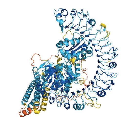
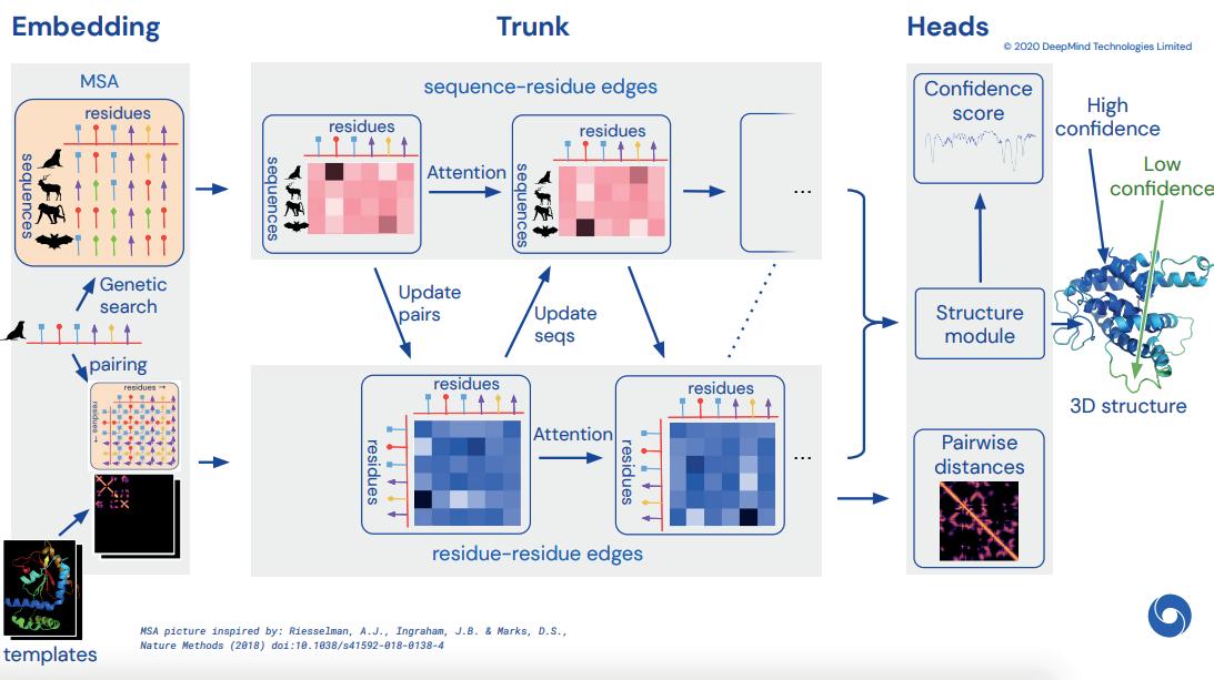
(Figure 6: How AlphaFold 2 works [13])
25 Physics and Technology
5. Conclusion
Ultimately, AlphaFold is undoubtedly a revolutionary computational method that can accelerate the process of discovering proteins exponentially, especially since 98.5% of full chain human proteins can be predicted by AlphaFold [8]. While it cannot replace existing experimental methods or solve the protein folding problem, it can act as a guide for scientists to use, providing them with hypotheses for the structure of a protein.
6. Bibliography
[1] Thompson, Michael C., et al. “Advances in Methods for Atomic Resolution Macromolecular Structure Determination.” F1000Research, F1000Research, 2 July 2020.https://f1000research.com/articles/9-667/v1
[2] Smyth, M S, and J H Martin. “X Ray Crystallography.” Molecular Pathology : MP, U.S. National Library of Medicine, Feb. 2000. https:// www.ncbi.nlm.nih.gov/pmc/articles/PMC1186895/
[3] Doerr, Allison. “Single-Particle Cryo-Electron Microscopy.” Nature News, Nature Publishing Group, 30 Dec. 2015. https://www.nature.com/ articles/nmeth.3700
[4] Zinkel, Brian. “What Is NMR Spectroscopy and How Does It Work?” Nanalysis, Nanalysis, 28 June 2019.https://www.nanalysis.com/ nmready-blog/2019/6/26/what-is-nmr-spectrography-and-how-does-it-work#:~:text=How%20Does%20NMR%20Actually%20Work,at%20 their%20own%20specific%20frequencies
[5] “Protein Data Bank: the Single Global Archive for 3D Macromolecular Structure Data.” Academic.oup.com, 8 Jan. 2019. https://academic. oup.com/nar/article/47/D1/D520/5144142
[6] Jaskolski, Mariusz, et al. A Brief History of Macromolecular Crystallography, Illustrated by a ... 3 Apr. 2014. https://febs.onlinelibrary.wiley. com/doi/10.1111/febs.12796
[7] Jumper, John, et al. “Highly Accurate Protein Structure Prediction with Alphafold.” Nature News, Nature Publishing Group, 15 July 2021. https://www.nature.com/articles/s41586-021-03819-2
[8] Tunyasuvunakool, Kathryn, et al. “Highly Accurate Protein Structure Prediction for the Human Proteome.” Nature News, Nature Publishing Group, 22 July 2021.https://www.nature.com/articles/s41586-021-03828-1
[9] Skolnick, Jeffrey, et al. “Alphafold 2: Why It Works and Its Implications for Understanding the Relationships of Protein Sequence, Structure, and Function.” Journal of Chemical Information and Modeling, U.S. National Library of Medicine, 25 Oct. 2021. https://www.ncbi.nlm.nih.gov/ pmc/articles/PMC8592092/
[10] Senior, Andrew W., et al. Protein Structure Prediction Using Multiple Deep ... - Wiley Online Library. 10 Oct. 2019. https://onlinelibrary. wiley.com/doi/full/10.1002/prot.25834
[11] Skolnick, Jeffrey, et al. “Alphafold 2: Why It Works and Its Implications for Understanding the Relationships of Protein Sequence, Structure, and Function.” Journal of Chemical Information and Modeling, U.S. National Library of Medicine, 25 Oct. 2021. https://www.ncbi.nlm.nih.gov/ pmc/articles/PMC8592092/
[12] Jumper, John, et al. Applying and Improving Alphafold at CASP14 - Wiley Online Library. 2 Oct. 2021. https://onlinelibrary.wiley.com/ doi/full/10.1002/prot.26257
[13] Jumper, John, et al. Alphafold 2 - Mimuw.edu.pl. 1 Dec. 2020https://www.mimuw.edu.pl/~lukaskoz/teaching/adp/lectures/lecture6/2020_12_01_TS_predictor_AlphaFold2.pdf
[14] Database, AlphaFold Protein Structure. “Alphafold FAQs.” Alphafold Protein Structure Database. https://alphafold.ebi.ac.uk/
[15] “Cryo-Electron Microscopy: Small Electrons to Visualize Large Molecules.” Università Vita-Salute San Raffaele, 16 June 2020, https://www. unisr.it/en/news/2020/6/criomicroscopia-elettronica-piccoli-elettroni-per-visualizzare-grandi-molecole.
[16] Example of a One-Dimensional NMR Spectrum of a Small Protein ... https://www.researchgate.net/figure/Example-of-a-one-dimensionalNMR-spectrum-of-a-small-protein-Rubredoxin-with_fig2_224830551.
26 Scientific Harrovian 2022

the FUNDAMENTALS of
QUANTUM COMPUTING
by Sen Yi Mok
27 Physics and Technology
1. Introduction
Estimated to be a one trillion dollar industry, quantum computing is a revolutionary new field which would reach market sizes close to the global tourism industry [1,2]. Instead of standard bits that store memory in supercomputers, quantum computers use qubits (also known as quantum bits) which can represent a huge number of states simultaneously [3]. Through the uses of qubits and quantum physics, quantum computing has been proven to be able to solve BQP (Bounded-error Quantum Polynomial) problems in the subset of NP (Nondeterministic polynomial) problems that typical supercomputers can never feasibly be able to solve [4,5]. This is also referred to as quantum supremacy. This would allow massive developments in quantum chemistry like ab initio calculations, encryption, weather forecasting, and stock market analysis [2,6].
Quantum supremacy has been first claimed to be proven in October 2019 by the 54-qubit processor Google “Sycamore” despite comments by IBM researchers that the supercomputer “Summit” could achieve similar results in 2.5 days [7,8]. Quantum supremacy was later demonstrated by China’s 113-qubit “Jiuzhang 2.0” in 2020 (1024 times faster than supercomputers) and currently, IBM’s 127-qubit “Eagle” developed in late 2021 is the fastest quantum computer as of 10 Nov 2022 [7,8].
2. Superposition
The reason why quantum computers are so much more powerful than regular supercomputers is because of two phenomena of quantum physics: superposition and entanglement [9]. Unlike regular bits that can only store ‘0’s or ‘1’s, qubits, a two-level quantum system can store a linear combination of basis states which can act like axes on a plane [10]. For qubits, the basis states are the ket-vectors : | 0 > and | 1 >. Similar to vectors, the superposition of qubits can be thought of as a combination of different magnitudes of the basis states or adding vectors together [10]. For example, a superposition of a qubit can be < | 0 > + <√3 | 1 >. The left- hand side (< | or < | ) is known as the bra-vectors and combined with the right ket-vectors ( 0 > or 1 > ) make up the bra-ket notation. It is key to note that this superposition is simply one of an infinite number of potential combinations of different magnitudes of vectors, but not multiple states at once [10]. The special property of superposition allows qubits to represent over 2n potential states at the same time with only n qubits [9].


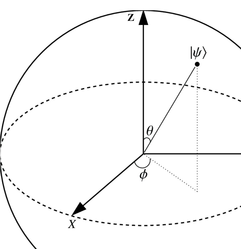
To represent the superposition of qubits, we can use 2 constants α and β which are complex numbers (for expressing wave functions of subatomic particles) where | > = | 0> + | 1> and |α|2 + |β|2 = 1 [11]. Despite having four components (real and imaginary parts of α and β), the 3D Bloch sphere can be used to visualise the points after simplifying the equation into: where (phi) is the imaginary part of β ( ) - the imaginary part of ( ) which represents the angle between the x-axis and where the point (psi) touches the XY plane whilst (theta) represents the angle from the z-axis and the line to the point from the origin [11]. Using the above equation, we can deduce that an arrow pointing up along the z-axis means that the qubit will always collapse into | 0 > and vice versa.


from


28 Scientific Harrovian 2022
Figure 1 shows a Bloch Sphere as described above [12].
However, when qubits are measured, they collapse into one of their eigenstates which is either | 0 > or | 1 > based on probabilities in the bra-ket notation.

Given the above example, < | 0 > + < | 1 >, to find the probability of the qubit that will converge into | 0 >, first square this expanded expression < 0 | 0 > + < 0 | 1 > which results in this expression < 0 | 0 > + < 0 | 1 >. It is important to note that < 0 | 0 > and < 1 | 1 > is equivalent to 1 as the bra-vector matches the ket-vector whilst the bra-vectors < 1 | 0 > and < 0 | 1 > is 0 as the two vectors are not equivalent [10]. Therefore, we get x 1 + x 0 which is equivalent to or 25%. On the other hand, you can find the probability that the qubit will collapse into | 1 > using the same method which results in 75% or you can subtract the probability of collapsing into the eigenstate | 0 > from 1 which would be 1 - in this case and leads to the same result: 75%.
3. Entanglement

Another property of qubits is entanglement. Entanglement, otherwise known as “spooky action at a distance” by Einstein, refers to the fact that a pair of particles can share a distinct feature where the measurement of the first particle in the pair is perfectly correlated with the second particle in the pair [9, 13]. For example, if two subatomic particles (Particle A and Particle B) are entangled and the total ‘spin’ of the system is 0, if Particle A is measured to be counterclockwise, Particle B is guaranteed to have a measurement that is clockwise instantaneously after Particle A’s measurement no matter how far the particles are from each other [13]. However, communication using these particles is impossible as it is impossible to determine their final state before measurement and there is no way to copy any of the particles [9, 37]. This is due to the no-cloning theorem, which means that it is impossible to abstract information about the two coefficients of the superposition [37]. Entanglement can help speed up quantum computers by using the determined properties of other entangled qubits and is shown to be necessary for quantum supremacy [14].
4. Quantum Gates
Quantum gates are used in quantum computing to make necessary calculations and can be represented by matrices. They can alter the states of the qubit when the gates are applied to the qubits and are always reversible [15]. Unlike regular logic gates like AND or NOT gates, the input and outputs of these quantum gates can be in superposition [16]. Here are a few examples of single qubit quantum gates:
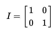
The Identity Gate, or the ‘I’ Gate, acts as a do-nothing operation and does not change anything about the qubit.
The Hadamard Gate essentially changes the state of the qubit so it is in a superposition such that it has an equal probability of converging to either the | 0 > or | 1 > eigenstate when observed [15]. It can also be described as a rotation around the Bloch sphere vector (1, 0, 1) [17].
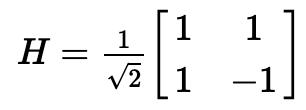 Figure 2 shows the Identity Matrix or the Identity Gate [17].
29 Physics and Technology
Figure 3 shows the Hadamard Gate Matrix [17].
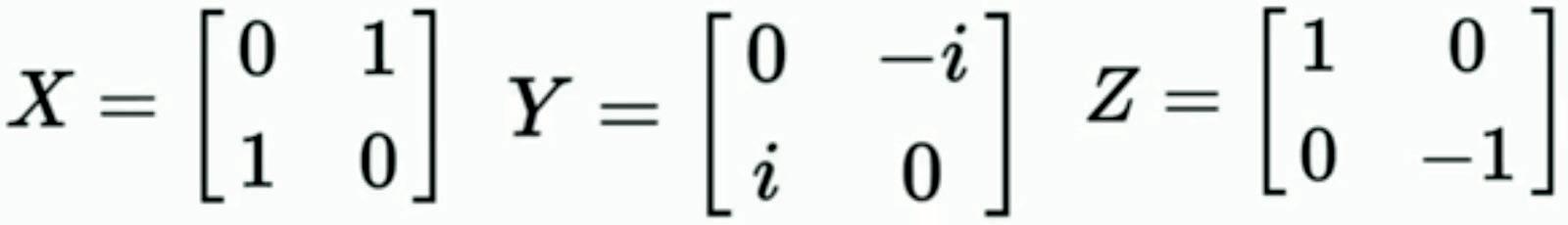
Pauli Gates, flips the qubit in the X, Y or Z axis depending on the specific Pauli Gate along their position on the Bloch Sphere. These gates are also known as X, Y or Z Gates. For example, after applying the X Pauli Gate (equivalent to the NOT gate in classical computers), the Z position is inverted whilst the X and Z positions are inverted after applying the Y Pauli Gate, and only the X position is inverted after applying the Z Pauli Gate [15, 17].
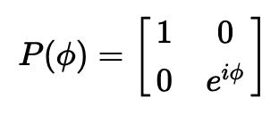
The Phase Shift Gate, or the P gate, takes a real number and rotates the qubit around the Z axis of the Bloch Sphere for radians [17]. The Z Gate is equivalent to P(π). To represent 90° degree turns around the Z axis of the Bloch Sphere, the S gate, or the Gate is used which is equivalent to P(π/2) [17]. Similarly, to represent a 45° degree turn, the T gate, or the Gate is used which is equivalent to P(π/4) and the inverse of the T gate, the gate is equivalent to P(-π/4) [17].


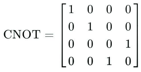
Some gates perform operations on two qubits. For example, the controlled-NOT gate or the CNOT gate inverts the other qubit if the indicator qubit is | 1 > [15]. For example, if the indicator qubit is | 1 > and the other qubit is | 0 >, the result would be | 1 1 >.
Another important widely used gate is the SWAP gate which swaps the values of the two qubits with each other [15]. For example, two qubits with states | 1 0 > will result in | 0 1 > after the SWAP gate is applied to it.
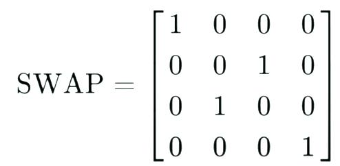 Figure 4 shows the 3 Pauli Gate Matrixes [15].
Figure 5 shows the Phase Shift Gate Matrix [17].
Figure 6 shows the CNOT Gate Matrix[15]
Figure 4 shows the 3 Pauli Gate Matrixes [15].
Figure 5 shows the Phase Shift Gate Matrix [17].
Figure 6 shows the CNOT Gate Matrix[15]
30 Scientific Harrovian 2022
Figure 7 shows the SWAP Gate Matrix [15].
Through the manipulation of quantum gates and using the properties of qubits, quantum computers are able to do complex calculations. However, since a qubit collapses to one of the eigenstates | 0 > or | 1 > based on probability, these calculations have to be repeated several times to ensure that the output matches the result that should be obtained [16].
5. Qubits
There are several different types of qubits and this article will be covering the three most common types: electrons in atoms or ions, photons, and superconducting circuits.
Subatomic particles, like electrons, have an inherent property known as spin, which is a type of angular momentum [18]. They naturally behave as if they are spinning and initially have a non-zero angular momentum despite not rotating around another object [18]. Thus using the property, we can find that the electron is either in the spin state ‘spin up’, if it is ‘rotating’ clockwise or the spin state ‘spin down’ if it is ‘rotating’ anticlockwise [18]. The spin states ‘spin up’ and ‘spin down’ corresponds to the | 0 > or | 1 > eigenstates [19]. We can also alter their energy state (switching between their natural state and their “excited” state) using lasers to represent the two eigenstates.
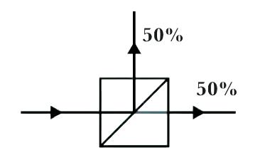
Photons, which are very small ‘packets’ of light, can also be used in several ways to model the two eigenstates [19]. Path qubits model the eigenstates by having a single photon pass through a beam splitter which has two light detectors on their respective sides. This causes either light detector to detect a photon 50% of the time but never at the same time, as the photon cannot be split [20].
The top light detector can represent a | 0 > state and the bottom light detector can represent a | 1 > state [19]. Another property of photons is that they have one of two polarisations (horizontal [H] and vertical [V]) perpendicular to the direction of wave propagation as light is a transverse wave which oscillates. These photons can also exist in a superposition of the two polarisations or have one of the two polarisations, thus they are known as polarisation qubits. Calculations can be made when passing these photons through a horizontal or vertical polariser. A horizontal polariser will let a photon with a horizontal polarisation through but not a photon with a vertical polarisation and vice versa for the vertical polariser [20]. The probability that a photon with a superposition can pass through the horizontal polariser will be |α|2 whilst the probability it will pass through the vertical polariser will be |β|2 [20].
Unlike classical computers where there are multiple electrons in a DRAM circuit for redundancy in case of electron leaks, qubits in quantum computers can be easily collapsed when they interact with other subatomic particles and struggle to work with bigger bits with more electrons [21]. However, this means that they would be vulnerable to electron leakage. A solution around this is superconducting circuits, which are certain metals that are cooled down to nearly 1°K so that their electrons are joined together as a unit and do not scatter around [21]. We can measure the energy level of the electrons or the direction of current to represent the two eigenstates of the qubit [19, 21].
Figure 8 shows a photon passing through a beam splitter[20]
31 Physics and Technology
6. Advantages
One of the most important features of quantum computers is their computational power and speed. Unlike supercomputers, quantum computers can double their computational speed by adding a single qubit [9]. Given that IBM released their 5-qubit quantum computer in May 2016 and has created a computer with 25.4 times more qubits in just over 5 years, the potential for quantum computers is huge [22]. They also have the ability to solve complex problems that have many interacting variables and run multiplex simulations due to superposition and entanglement [23]. For example, Quantum Computing Inc (QCI) has designed quantum computers which have been used to solve a 3854-variable optimization problem with 500 constraints for placing vehicle sensors in a BMW in under six minutes and they were able to find a solution with 96% vehicle coverage with only 15 sensors [24]. Aside from cars, quantum computers can be used in other fields for cryptography, chemistry, medical usage, modelling, forecasting, and much more [6, 23]. Quantum computers also have a lower lower bound than classical computers at regular processes like ordered searching [ log (n) compared to log (n)], comparison-based sorting [O(n) compared to O(n log n)] and element distinctiveness [O(√n) compared to O(n log n)[25] In addition to these advantages, quantum computing is also environmentally friendly and requires only 0.002% of the energy used by a classical computer [26].
7. Disadvantages
Although quantum computers can be used in a variety of different fields, they are unable to solve most problems with 100% accuracy that can be solved with classical computers easily due to their probabilistic nature [16]. Quantum computers are also unable to store data and most memory can only be stored for up to a few hundred microseconds (10-6 s).
To reduce errors in qubits, they also have to be stored in extremely cold temperatures (3 °K) and require huge machinery as well as energy [27]. Although there have been many recent advancements, we are still far away from unleashing the full potential of quantum computing as quantum computers with hundreds or thousands of qubits are extremely complicated and difficult to build as qubits tend to have lower connectivity (communication within qubits) in larger quantum machines [27]. Due to its technical limitations, the difficulty of building a supercomputer and the extreme environment needed to store a quantum computer, a McKinsey report predicts that there will still be less than 5000 quantum computers by 2030, compared to over 2 billion computers today [27]. However, the rise of quantum computing means that many companies will now be at risk of being hacked by quantum computers as they will be able to break current cryptographic algorithms with ease [28]. They are also very expensive and cost tens of millions of dollars for one.
8. Cryptography
Currently, there have been multiple quantum computing algorithms such as Shor’s algorithm and Grover’s algorithm which have been developed to crack encryptions such as RSA (involves multiplying two huge prime numbers together), TDES or AES. Shor’s algorithm helps decrypt encryptions like RSA as it is able to find the decomposition of any integer into two primes in O(d3), where d is the number of digits that the integer has in decimal, which is a massive speedup compared to the exponential time complexity of classical computers [29].This time is very short considering that the upper bound of RSA integers is around 2470 digits long. Grover’s algorithm can help crack symmetric key algorithms with a lower number of bits. Using quantum computation to search for elements in an unstructured database allows for an O(√n) time complexity compared to O(n) in classical computers [30]. Although this is only a quadratic speed-up, this is already enough to crack any key size of TDES and up to 128-bit AES [30, 31]. However, quantum computing is still in its infancy stage and quantum scientists have not been able to use these algorithms to decrypt huge numbers currently used today. Despite this, many companies have invested in quantum-safe algorithms like CRYSTALS-Kyber, CRYSTALS-Dilithium and Falcon [31].
32 Scientific Harrovian 2022
8. Chemistry
In quantum chemistry, ab initio (“from first principles”) calculations try to solve the electronic Schrödinger equation, = , given the position of the nuclei and number of electrons to find its energy and wave function, which can be derived to find electron densities, electron distribution and any other properties of the system [32]. As the calculations are very complex and are probabilistic, quantum computers have been used to represent states of a quantum chemical system to simulate quantum physics. Although current quantum computers have a relatively low number of qubits and have limited gate operations, future quantum computers with more qubits will be able to run a quantum phase estimation (QPE) algorithm which can solve for any variables in polynomial time, a time that is unreachable for classical computers [33]. Currently, quantum computers by IBM have been successful in doing some ab initio molecular dynamic methods (simulation of physical movement of subatomic particles) with fairly high precision on simpler elements like hydrogen and even beryllium hydride [33, 34].

9. Prediction
Weather forecasting, stock market analysis, and predictions are all very complex and take lots of computational power to get an accurate prediction. These can be solved through quantum computing, as properties of superposition allow for the handling of huge numbers of variables interacting in a non-trivial way, which can reduce the damages of natural disasters as the predictions will become more accurate and precise [35]. Through the use of qubits, quantum machine learning algorithms can also be developed for pattern recognition with huge datasets and performing classification of data [35]. This could help increase investment gains, open new investment opportunities and reduce the risk of trading [36]. Additionally, it can help detect fraud (over $10 billion is lost per year due to fraud in the US), money laundering and forecast crashes in the markets which can save billions and billions of dollars [36]. Quantum computers can also help with recommender systems and social media algorithms.
10. Conclusion
Despite the extreme conditions, expenses, and issues raised due to quantum computing such as privacy concerns, quantum computing will surely become one of the most important industries in the world within a decade due to its strong ability to solve very hard optimization problems in a short amount of time. Although quantum computing is still in its infancy, its applications in so many different fields like cryptography, chemistry and forecasting still shock many and are full of potential. Through further research by scientists, I believe that quantum computers with thousands of qubits, which can perform P or even NP problems in very little time, can be made in ten to fifteen years given the exponential growth of quantum computing technology and rising awareness surrounding this technology.
*Note that this article does not cover more advanced topics such as quantum interference or quantum algorithms or applications of quantum gates to make this more simple and digestible for the reader.
33 Physics and Technology
11. Bibliography
[1] “Global Tourism - Industry Data, Trends, Stats | IBISWorld.” IBISWorld - Industry Market Research, Reports, & Statistics, https://www.ibisworld.com/global/ market-research-reports/global-tourism-industry/.
[2] “Quantum Computing Is Coming. What Can It Do?” Harvard Business Review, https://www.facebook.com/HBR, 16 July 2021, https://hbr.org/2021/07/quantum-computing-is-coming-what-can-it-do.
[3] Lu, Donna. “What Is a Quantum Computer?” NewScientist, https://www.newscientist.com/question/what-is-a-quantum-computer/.
[4] “Google AI Blog: Quantum Supremacy Using a Programmable Superconducting Processor.” Google AI Blog, https://ai.googleblog.com/2019/10/quantum-supremacy-using-programmable.html.
[5] Aaronson, Scott. “The Limits of Quantum.” SciAm, SpringerNature, 2008, https://www.cs.virginia.edu/~robins/The_Limits_of_Quantum_Computers.pdf.
[6] Bellapu, Apoorva. “10 Difficult Problems Quantum Computers Can Solve Easily.” AnalyticsInsight, https://www.analyticsinsight.net/10-difficult-problems-quantum-computers-can-solve-easily/.
[7] Choi, Charles Q. “Two of World’s Biggest Quantum Computers Made in China - IEEE Spectrum.” IEEE Spectrum, IEEE Spectrum, 6 Nov. 2021, https://spectrum.ieee.org/quantum-computing-china.
[8] “IBM Unleashes the Eagle, the World’s Most Powerful Quantum Processor.” New Atlas, 17 Nov. 2021, https://newatlas.com/quantum-computing/ibm-eagle-quantum-processor/.
[9] Voorhoede, De. “Superposition and Entanglement.” Quantum Inspire, https://www.quantum-inspire.com/kbase/superposition-and-entanglement/.
[10] Quantum Superposition, Explained Without Woo Woo, YouTube, uploaded by TheScienceAsylum, 29 Nov 2021, https://www.youtube.com/watch?v=ZUipVyVOm-Y
[11] The Bloch Sphere (simply explained), YouTube, uploaded by mu-hoch-3, 11 Jun 2020, https://www.youtube.com/watch?v=a-dIl1Y1aTs
[12] Contextual Semantics: From Quantum Mechanics to Logic, Databases, Constraints, and Complexity - Scientific Figure on ResearchGate.
[13] Quantum Computers: Superposition, Entanglement, and Qubit, Youtube, uploaded by ScienceyStuff, 9 Apr 2020, https://www.youtube.com/watch?v=x3LmPFSZAAU
[14] Is entanglement the key to quantum computing?, Youtube, uploaded by LookingGlassUniverse, 8 May 2021, https://www.youtube.com/watch?v=4RTxJ_I9LtU
[15] Quantum Gates, Youtube, uploaded by Travis Gritter, 6 Feb 2017, https://www.youtube.com/watch?v=gz5rjhiU4ao
[16] Quantum Computers Explained - Limits of Human Technology, Youtube, uploaded by Kurzgesagt - In a Nutshell, 8 Dec 2015, https://www.youtube.com/ watch?v=JhHMJCUmq28
[17] The Qiskit Team. “Single Qubit Gates.” Qiskit.Org, Data 100 at UC Berkeley, 6 July 2022, https://qiskit.org/textbook/ch-states/single-qubit-gates.html.
[18] Spin in Quantum Mechanics: What Is It and Why Are Electrons Spin 1/2? Physics Basics, Youtube, uploaded by Parth G, 4 Nov 2020, https://www.youtube. com/watch?v=DCrvanB2UWA
[19] “What Is a Qubit? | Institute for Quantum Computing.” University of Waterloo, https://uwaterloo.ca/institute-for-quantum-computing/quantum-101/quantum-information-science-and-technology/what-qubit
[20] Quantum Mechanics 2 - Optical Qubits: Polarisation and Interference, Youtube, uploaded by Centre for Quantum Technologies, 12 Oct 2021, https://www. youtube.com/watch?v=sTxQZcTSw-4
[21] Building a quantum computer with superconducting qubits (QuantumCasts), Youtube, uploaded by TensorFlow, 8 Feb 2019, https://www.youtube.com/ watch?v=uPw9nkJAwDY
[22] “Five Experimental Tests on the 5-Qubit IBM Quantum Computer.” SCIRP Open Access, https://www.scirp.org/journal/paperinformation.aspx?paperid=86139.
[23] FutureLearn. “What Is Quantum Computing? Essential Concepts and Uses - FutureLearn.” FutureLearn, https://www.facebook.com/FutureLearn, 15 Oct. 2021, https://www.futurelearn.com/info/blog/what-is-quantum-computing.
[24] Pires, Francisco. “BMW’s 3,854-Variable Problem Solved in Six Minutes With Quantum Computing | Tom’s Hardware.” Tom’s Hardware, Tom’s Hardware, 28 July 2022, https://www.tomshardware.com/news/quantum-computing-company-solves-3854-variable-problem-for-bmw-in-six-minutes.
[25] Hoyer, Peter, et al. “Quantum Complexities of Ordered Searching, Sorting and Element Distinctness.” ArXiv, 15 Feb. 2001, https://arxiv.org/abs/quantph/0102078.
[26]Wu, Tin Lok. “What Is Quantum Computing and How Can It Help Mitigate Climate Change? | Earth.Org.” Earth.Org, Earth.Org, 22 Aug. 2022, https:// earth.org/what-is-quantum-computing/.
[27] “Will Quantum Computing Replace Traditional Methods? | Built In.” Built In, https://builtin.com/software-engineering-perspectives/quantum-classical-computing.
[28] “The Impact of Quantum Computing on Society | Post Quantum Cryptography | DigiCert.” SSL Digital Certificate Authority | Encryption & Authentication | DigiCert.Com, https://www.digicert.com/blog/the-impact-of-quantum-computing-on-society.
[29] “Shor’s Algorithm - IBM Quantum” IBM Quantum, https://quantum-computing.ibm.com/composer/docs/iqx/guide/shors-algorithm.
[30] Mina-Zicu, M.; Simion, E. Threats to Modern Cryptography: Grover’s Algorithm. Preprints 2020, 2020090677
[31] “What Is Quantum-Safe Cryptography, and Why Do We Need It? | IBM.” IBM - United States, https://www.ibm.com/cloud/blog/what-is-quantum-safe-cryptography-and-why-do-we-need-it.
[32] Cyanide, Mohsin. Ab Initio Calculations and Modelling in Computational Chemistry. YouTube, 9 Jan. 2022, https://www.youtube.com/watch?v=LRK0zgNjPl8.
[33] Fedorov, Dmitry, et al. “Ab Initio Molecular Dynamics on Quantum Computers.” ArXiv.Org, 14 Aug. 2020, https://arxiv.org/abs/2008.06562.
[34]“Science | AAAS.” AAAS, https://www.science.org/content/article/quantum-computer-simulates-largest-molecule-yet-sparking-hope-future-drug-discoveries?cookieSet=1.
[35] Dutta, Aratrika. “Quantum Predictions: Weather Forecasting with Quantum Computers.” Analytics Insight, 27 Sep. 2021, https://www.analyticsinsight.net/ quantum-predictions-weather-forecasting-with-quantum-computers/.
[36] “Quantum Computing Use Cases for Financial Services | IBM.” IBM, https://www.ibm.com/thought-leadership/institute-business-value/report/exploring-quantum-financial.
[37] “The No-Cloning Theorem | Quantiki.” Quantiki | Quantum Information Portal and Wiki, https://www.quantiki.org/wiki/no-cloning-theorem.
34 Scientific Harrovian 2022

the SPEED OF LIGHT
and its significance
by Sky Lee
35 Physics and Technology
1. The Universal Threshold
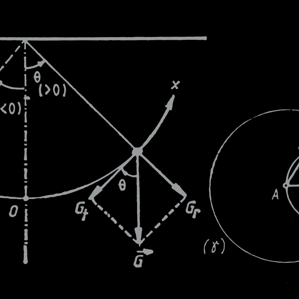
Would you believe me if I told you that the fastest speed in the vast universe is 299 792 458 ms-1, the speed of light, [1] that if something goes faster than that threshold, the universe breaks and reality collapses? The fact that the universe has a speed limit is extremely counterintuitive; it’s hard to picture something travelling at the speed of light and it’s even harder to grasp why nothing can go over that specific limit.
2. Origins
1905 was Einstein’s Annus Mirabilis, his Year of Miracles, [2] in which he published four papers: “On a Heuristic Viewpoint Concerning the Production and Transformation of Light”, “On the Motion of Small Particles Suspended in a Stationary Liquid, as Required by the Molecular Kinetic Theory of Heat”, “On the Electrodynamics of Moving Bodies”, and “Does the Inertia of a Body Depend Upon Its Energy Content?”. These four papers have made a significant impact on the physics community, giving insights in the Quantum Theory of Light and the existence of atoms and molecules. [3] Out of the four papers, “On the Electrodynamics of Moving Bodies” is probably the one that stands out most. Why, you may ask, because it includes the most famous equation in the world, E=mc2, the embodiment of the renowned Special Relativity and is the key to understanding how this universe operates.
3. The Speed of Light as an Invariant
Einstein’s Special Relativity shows the connection between some of the most significant quantities in the universe, mass, time, and space without the complication of gravity (relativity considering gravity is known as General Relativity). [4] Special relativity is based on the fact that the speed of light is a constant for all observers when gravity is not taken into consideration (curved light gets slowed down when it is not observed from one specific local reference frame according to the Shapiro Time Delay, assuming the presence of gravity) (fig A). [5] The constant c was calculated by Scottish physicist James Clerk Maxwell with the following equation (fig B).

With the fact that the speed of light is constant in mind, we can move on to understanding Special Relativity with the ‘spaceship and planet’ analogy.
fig. A Shapiro time delay [5]
36 Scientific Harrovian 2022
fig. B
4. Spaceship and Planet Analogy [6]
Consider a spaceship moving relative to a hypothetically stationary planet (fig. C), according to Einstein’s First Postulate of Special Relativity, the laws of physics are the same and can be stated in their simplest form in all inertial frames of reference [7].Thus, in this situation there is no way we can determine whether the planet is stationary, and the plane is moving or vice versa. Now imagine a ball being thrown across in the spaceship, the relative speed of the ball as observed from the planet, according to the Galilean Transformation, is the speed of impulsion of the ball combined with the speed of the spaceship itself. On the other hand, the relative speed of the ball as observed locally in the spaceship is just the speed of impulsion of the ball. With that said, the relative speed of the ball is lower if observed locally in the spaceship.
Instead of a ball being thrown in the spaceship, imagine a beam of light being shone across in the spaceship (fig. D). Intuitively, we would consider the beam of light as a travelling particle, just like the ball, and thus, assume that the relative speed of the beam of light is lower if observed locally in the spaceship. Yet, that contradicts Maxwell’s calculations of a constant speed of light c, for every observer, when gravity is not taken into consideration. So, is the speed of light c, still a constant? If it is, then classical Newtonian mechanics would simply be paradoxical. This is where special relativity comes in, a scientific principle which accommodates Maxwell’s constant speed of light and the validity of classical mechanics.

 fig. D A beam of light being shone across the spaceship [6]
37 Physics and Technology
fig. C A moving spaceship relative to a planet [6]
5. Mass
There are so many reasons why nothing can go faster than the speed of light. With that said, there is only one particular physical phenomenon that explains why only massless objects like photons can travel at the speed of light, that is Relativistic Kinetic Energy.
Rest mass energy is the energy (fig. E) that all matter possesses (i.e., as long as it has mass and takes up space), be it matter that is stationary or matter that has motion. Relativistic energy (fig. F) is the energy that every moving object possesses. Relativistic kinetic energy (fig. G) is the energy every object possesses strictly due to their motion (i.e., the object’s rest mass energy is excluded). What is the significance of that, you may ask. As seen in the relativistic kinetic energy equation, as the speed of the object v increases, v2 also increases, as v approaches c, the Lorentz Factor, γ tends to infinity. This implies that if an object has to travel at the speed of light, the required energy tends to infinity (fig. I). As it is impossible to supply an infinite amount of energy to anything, it is simply unfeasible for matter to travel at the speed of light.
38 Scientific Harrovian 2022
fig. I graph of Kinetic energy against Speed [9]
6. Side Effect of Travelling at c: Time Dilation
Imagine a person travelling near the speed of light, Alice, and a stationary person. They experience time differently according to time dilation in Einstein’s Special Relativity.
If Alice is travelling in a spaceship at the speed of 0.9c, 90% the speed of light, then it would take her 1 second to travel a distance of 0.9c metres (ignoring the effects of relativistic kinetic energy). In this case, T, the time in Alice’s frame of reference is 1 second. Yet to stationary observer Bob, it takes Alice 2.29 seconds to travel a distance of 0.9c metres according to the Time Dilation Equation (fig. J).
What is more fascinating is that Alice or Bob could technically be the stationary observer in this case according to Einstein’s First Postulate of Special Relativity. In Alice’s frame of reference, she is stationary and Bob is moving at 0.9c, while in Bob’s frame of reference, he is stationary and Alice is moving at 0.9c. This implies that to Bob, time flows slower in his frame than in Alice’s frame. While to Alice, time flows slower in her frame than in Bob’s frame.
7. Breaking Causality and the Emergence of Time Paradoxes, a Result of Time Dilation
Cause has to come before effect in Physics. Person A would never receive a message before Person B sends it to him, your table never gets wet before you spill the water, and a person’s head wouldn’t explode before a bullet hits him. Causality governs how the Universe operates, it is that one rule which cannot be broken.
Imagine your friend, Jonathan, receiving your text message before you’ve even pressed the ‘send’ button, that’s crazy!
If an object travels faster than the speed of light, causality would be broken, or in other words, the Universe goes nuts. There is not much mathematical proof on how going faster than the speed of light breaks causality, a simple analogy and example can perfectly illustrate the correlation.
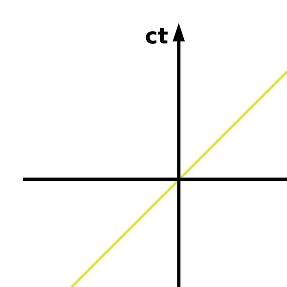

Before we move on to the example, we first have to understand Spacetime Diagrams. For every object, the horizontal line represents the change in space in its frame of reference while the vertical line represents the change of time in its frame of reference. For a stationary object, a vertical line represents its activity in spacetime. fig. K Spacetime Diagram [10]

39 Physics and Technology
Continuing with the example of Alice and Bob [11]. Imagine in a new scenario, Alice standing stationary on planet Earth and Bob moving at 0.87c away from Earth relative to her. In Alice’s frame of reference, she can be represented by a vertical line while Bob can be represented by a line of slope magnitude greater than 1. In Bob’s frame of reference, he can be represented by a vertical line while Alice can be represented by a line of slope magnitude greater than 1. Again, Einstein’s First Postulate of Special Relativity tells us that Alice can be stationary in her own frame of reference (fig. L) while Bob can also be stationary in his (fig. M). According to the Time Dilation Equation, in Alice’s frame of reference, 2 seconds for her is 1 second for Bob while in Bob’s frame of reference, 2 seconds for him is 1 second for Alice.
Enter FTL, Faster Than Light. Imagine there was something much faster than light, something that is instantaneous. An object travelling instantaneously can be represented as a horizontal line in spacetime diagram as an object travels an infinite distance in space without travelling in time. Imagine Alice sending an instantaneous message to Bob. If she sends the message to Bob, at 4 seconds on her clock (i.e. in her frame of reference), then Bob would receive her message at 2 seconds on his clock, if we consider this situation in Alice’s frame of reference (fig. N). Things start to get weird when we look at the message from Bob’s frame of reference, we can see that the message is sent from 4 seconds on Alice’s clock to 2 seconds on Bob’s clock, in other words, the message is sent from 8 second on Bob’s clock to 2 seconds on the same clock, the message has travelled back in time (represented by a negative slope) (fig. O) in Bob’s frame of reference! Now imagine Bob taking two seconds to read the message Alice sent him, and at 4 seconds on his clock, decides to send an instantaneous reply back to Alice. She would receive it at 2 seconds on her clock, meaning that she received a reply from Bob even before she sent the message (i.e. at 4 seconds on her clock in her frame of reference). Causality has been broken (fig. P) because of special relativity and the type of messaging system that can travel faster than the speed of light, in this example, at instantaneous speed.
fig. L Alice’s frame of reference [11]
40 Scientific Harrovian 2022
fig. M Bob’s frame of reference [11]
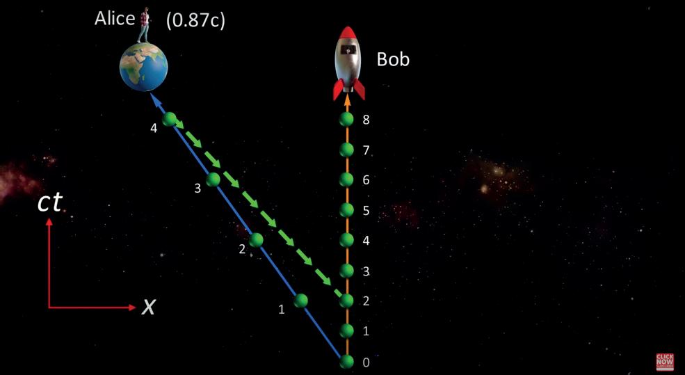
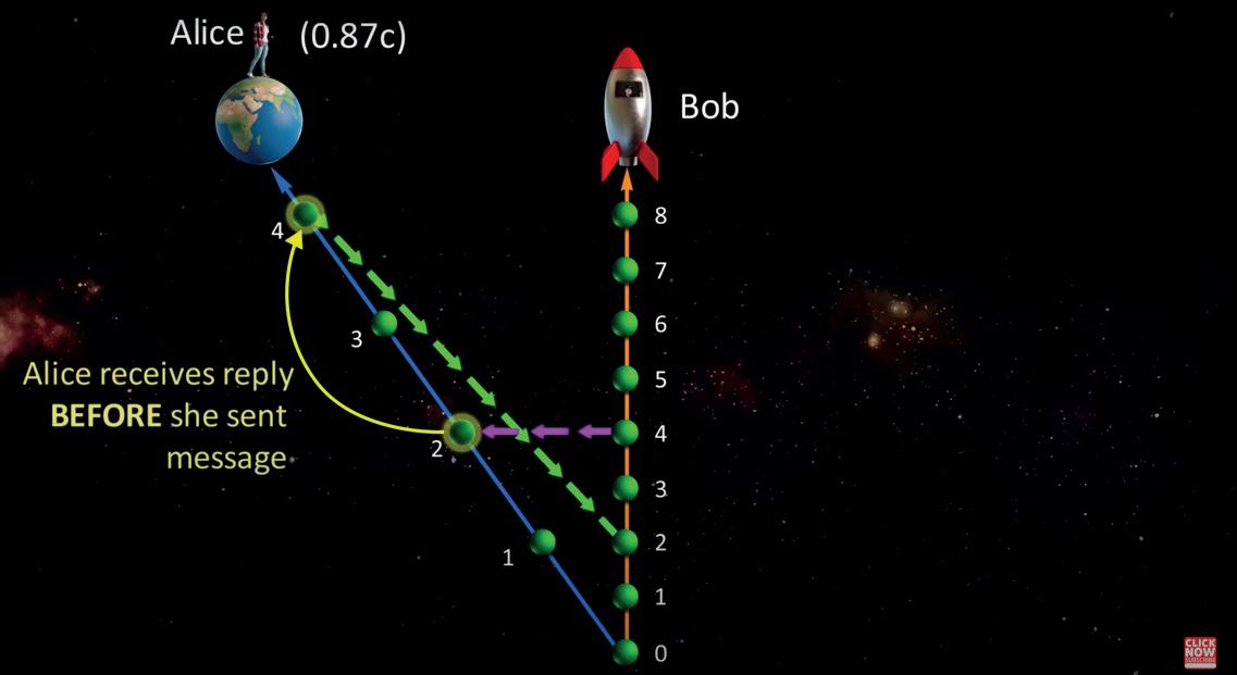 fig. N Alice’s frame of reference [11]
fig. O Alice’s message travelling back in time in Bob’s frame or reference [11]
fig. N Alice’s frame of reference [11]
fig. O Alice’s message travelling back in time in Bob’s frame or reference [11]
41 Physics and Technology
fig. P Causality has been broken
8. Insights
The speed of light is so much more than just a number used in calculations in physics tests, it is a value that governs how the universe operates and ensures effect comes after cause. It explains why you will never get a reply before you send a message, as there is nothing that can travel faster than the speed of light in this universe. While popular movies these days have explored the ideas of time travelling and travelling at the speed of light, the current physical laws remain unchanged, telling us concrete and undeniable facts about the universe: travelling quicker than the speed of light just isn’t feasible. ‘First comes the Physics, then comes the Engineering’, should humans endeavour on travelling at the speed of light, the fundamental laws of physics established by the great scientist, Einstein will have to be broken and overthrown.
9. Bibliography
[1] Wikipedia, “Speed of light”, accessed 2022 November 9, https://en.wikipedia.org/wiki/Speed_of_light
[2] npr, Richard Harris, “Albert Einstein’s Year of Miracles: Light Theory”, 2005, March 17, accessed 2022 November 9, https://www.npr. org/2005/03/17/4538324/albert-einsteins-year-of-miracles-light-theory#:~:text=Scientists%20call%201905%20Albert%20Einstein’s,the%20 famous%20equation%20E%3Dmc%C2%B2.
[3] Wikipedia, “Annus Mirabilis papers”, accessed 2022 November 9, https://en.wikipedia.org/wiki/Annus_mirabilis_papers#:~:text=The%20 annus%20mirabilis%20papers%20(from,the%20foundation%20of%20modern%20physics.
[4] Space, Vicky Stein, “Einstein’s Theory of Special Relativity”, 2021 September 21, accessed 2022 November 9, https://www.space. com/36273-theory-special-relativity.html#:~:text=Special%20relativity%20is%20an%20explanation,equation%20E%20%3D%20mc%5E2.
[5] Universerio@YouTube, “What is the true meaning of constant speed of light? Why is the Speed of Light Constant?”, 2022 August 22, accessed 2022 November 9, https://youtu.be/hvMAT1xeraM
[6] ScienceClic English@YouTube, “Special Relativity”, 2019 September 10, accessed 2022 November 9, https://www.youtube.com/watch?v=uTyAI1LbdgA
[7] Douglas College Physics 1207, 13.1 Einstein’s Postulates, no date, accessed 2022 November 9, https://pressbooks.bccampus.ca/introductorygeneralphysics2phys1207opticsfirst/chapter/28-1-einsteins-postulates/#:~:text=The%20first%20postulate%20of%20special,relative%20motion%20 of%20the%20source.
[8] For the Love of Physics@YouTube, “Relativistic Kinetic Energy | Answer to why nothing can travel faster than the speed of light?”, 2021 July 3, accessed 2022 November 9, https://www.youtube.com/watch?v=TwKSGVKfIXw
[9] LibreTexts Physics, “Relativistic Quantities”, 2020 November 6, accessed 2022 November 10, https://phys.libretexts.org/Bookshelves/University_Physics/Book%3A_Physics_(Boundless)/27%3A__Special_Relativity/27.3%3A_Relativistic_Quantities
[10] Wikipedia, “Spacetime Diagram”, accessed 2022 November 20, https://en.wikipedia.org/wiki/Spacetime_diagram
[11] Arvin Ash@YouTube, “How Faster than Light Speed Breaks CAUSALITY and creates Paradoxes”, 2021 June 25, accessed 2022 November 20, https://www.youtube.com/watch?v=mTf4eqdQXpA&t=744s
42 Scientific Harrovian 2022

STARLINK SATELLITE
Probability Collision Simulation
Based on Simplified Geometry Model
by Toby Li
43 Physics and Technology
Abstract
In this paper, a model based on a simplified geometry is introduced to give a very conservative collision probability prediction for the Starlink satellite in its most densely clustered region. Under the model in this paper, the probability of collision for Starlink satellite where it clustered most densely is found to be 8.484 * 10−4. It is found that the predicted collision probability increased nonlinearly with the increased safety distance set. This simple model provides evidence that the continuous development of maneuver avoidance systems is necessary for the future of the orbital safety of satellites under the harsher Lower Earth Orbit environment.
Introduction
With the advancement of modern rocket technology, more and more satellites are sent to Low Earth Orbital (LEO) to operate various missions. Satellites have taken an essential role in human society in different fields. Since the first artificial satellite was launched in 1957, a steady stream of satellite launches has increased space debris. For instance, in agriculture, the detection of crop growth situations and inspection of particular commercial crops. Regarding transport, satellites can monitor an urban traffic flow in peak periods and navigate for all car users. Until 30th April 2022, there were about 5,465 satellites in space functioning; out of them, 4,700 satellites orbit in LEO [1]. As one of the largest commercial satellite companies, SpaceX owns over 3300 Starlink satellites in LEO [2]. While the Federal Communications Commission (FCC) only permits launching up to 12,000 satellites, Space X planned to launch about 42,000 satellites in LEO [3]. However, with the increased number of satellites, space debris also increased significantly. This increases the probability of collisions in LEO and threatens all the satellites. According to the ESA’s MASTER-8 model, there are 36,500 objects larger than 10 cm and over 1.3 million between 1mm and 1cm in size in the current Earth orbit [4]. This numerous number of debris seriously endangers the safety of the existing operating satellite in every Earth orbit. There are several leading causes of space debris formation. About 39.52% of current space debris is generated due to the explosion of undisposed fuel on inactivated satellite or rocket components. Intentional breaks up of satellites make up 23.77% of total space debris, and several collisions observed contribute about 9.01% [5] of current Space debris. Fig (1) shows that the number of tracked space debris has increased rapidly since 1960 and poses a significant risk to satellites. From Fig (1), the overall trend of debris numbers is increasing, and an increasing trend can be observed with the recent year’s data [6]
44 Scientific Harrovian 2022
Figure 1. Number of debris observed since 1960
1.1 Problem Statement
The rapid increase in the number of space junk increased the probability of collisions of commercial satellites with various space debris. For instance, on 2009 February 10, the commercial satellite Iridium-33 and the Cosmos-2251, a decommissioned Russian military spacecraft, collided. Over 2201 debris fragments [7] were tracked from then until June of 2012, and clouds of the fragment were detected after the collision, as shown in Fig (2). It is predicted that the debris from Cosmos-2251 will stay in orbit for over the next 20–30 years and that from Iridium 33 for over 100 years in LEO.
Although the collision probability is almost zero after each country regulates the satellite, the collision probability still exists when the satellite encounters random space junk. Faced with this situation, the satellite needs to perform maneuver avoidance to reduce the risk of collision [8], yet this will consume the satellite’s fuel and reduce the satellite’s lifetime in orbit. Therefore, it is significant to effectively predict the collision probability and adjust in advance for the on-orbit life of satellites and the space safety of LEO. Especially for Starlink satellites, which are one of the largest satellite collections, collision probability prediction is more necessary. One of the recent papers published in 2022 focuses on Starlink satellite in-orbit secondary collision risk using a matrix, integration, and MASTER-8 model to predict the probability of collision between Starlink satellite and debris [9]. This paper focuses on predicting the collision probability of Starlink satellites at the most densely clustered altitude in LEO. However, for LEO collision, there still lack of research focused on Starlink collision.
1.2 Model Formulation
A conventional method used in the past to examine the necessity of maneuver avoidance is whenever a tracked object is expected to cross a keep-out zone of 5 * 2 * 2 km centered on the Orbiter. However, this method does not predict collisions probability between debris and satellite. Significant progress has been made in the collision probability calculation problem that some researchers have been working to formulate and computerize. At first, researchers deal with three-dimensional probability density. To reduce computation, research assumes the satellite to be spherical. To obtain a two-dimensional integral over a circular region. In 2001, Russell P. Patera, instead of integrating over an area, one integrates around its edge to speed up processing by lowering the number of times the integrand must be evaluated [10]. This method does not need to assume a spherical shape, leading to more accurate results. Inspired by this research, this paper reduces three-dimensional space into two-dimensional space by projecting a cross-sectional area around the circular path.
Instead of using a complex series of calculus and matrices to predict the probability of collision, this paper takes a spatial geometrically based approach to predict a very conservative collision probability. Although the method is relatively coarse compared to existing methods, it does not give a very accurate prediction of collision probability. However, this model can give a rough estimate of the collision probability in a short time because the model does not require complex computation and data. With the advantage of using the geometric properties of the space for calculation, the model in this paper can be better understood by some non-experts unfamiliar with the field.
45 Physics and Technology
Figure 2. The trajectory of debris created by the collision (Direct reference of pictures approved by the original author)
1.3 Section Review
This paper aims to investigate the collision probability in regions with a high density of Starlink satellites. Specifically, focus on the LEO region where Starlink is distributed most densely. There are two scenarios; the first scenario investigates the Starlink satellite’s collision probability with debris in 2022 at an altitude of 539-561km. The second scenario predicts the average collision probability of Starlink satellites between 2022 to 2040 at an altitude of 525-535km, assuming there are 10080 Satellites in the region described. In section 2, a systematic analysis of the environment that is investigated is introduced. The factors that are significant for the research, such as the distribution of the satellite and prediction of the number of debris, are discussed in the section. section 3 focuses on the modeling; a probability model based on the Monte-Carlo method is used to approximate the collision probability between Starlink Satellite and debris at the selected altitudes. The section mainly describes how to gradually simplify a physical problem and convert it into a mathematical probability problem while considering a certain accuracy. Section 4 mainly analyzes the experimental results of the model. In this section, the uncertainty caused by model simplification is also discussed, and an analysis of the uncertainty is given. Finally, section 5 reviews the analysis and experiments conducted throughout the paper and evaluates the overall advantages and disadvantages of the model. The effectiveness of improving the model to the collision probability is also discussed.
System Analysis
2.1 Space Environment Introduction
In this paper, a standard from the National Aeronautics and Space Administration (NASA) is used for the division of Earth satellites. That is the altitude from 160km-2000 km. The Earth radius chosen in this paper is the equatorial radius of Earth, which is 6378.13 km. This paper mainly focuses on the distribution of SpaceX satellites and debris in LEO. Debris and satellite density are calculated for the dataset set up in this section.
2.2 Debris Distribution
This paper only focuses on some crucial information that affects the prediction of collision of probability: launched year and decay year of debris and apogee and perigee of debris. The debris data is from the United States Space Command (USSPACECOM) [6], which includes all the information about the debris that has appeared in LEO since 1957. The total debris number is the sum of debris and rocket bodies. In 2022, 14,825 pieces of debris were observed in LEO, and 3,032 pieces of debris are in the region of 539km-561km. Since the debris trajectory is uncertain and random, it is assumed in this paper that the debris in LEO is uniformly distributed. Therefore, the density of LEO debris can be derived from the density formula. Debris density at the altitude of 539-561km can be obtained through the following formula. In the formula, Ndebris is the number of satellites in the selected region, REarth is the radius of Earth, Hhigh is the altitude of higher of the selected region, and Hlow is the altitude of lower of the selected region.
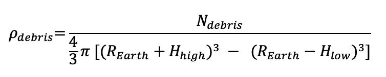
From the dataset collected, debris density in 539km -561km calculated is 2.285*10-7 debris/km3.To model the debris density in the future, the number of debris in consecutive years in the short future must be predicted, and this prediction is based on the number of debris tracked in the past since the first rocket was launched. Following is the debris dataset that tracked objects at an altitude of 525-535km since 1957; prediction for debris numbers in the future is based on these data.
According to Fig (3), the trend of the number of debris can be observed from 1957 to 2022. An interpola- 46 Scientific Harrovian 2022
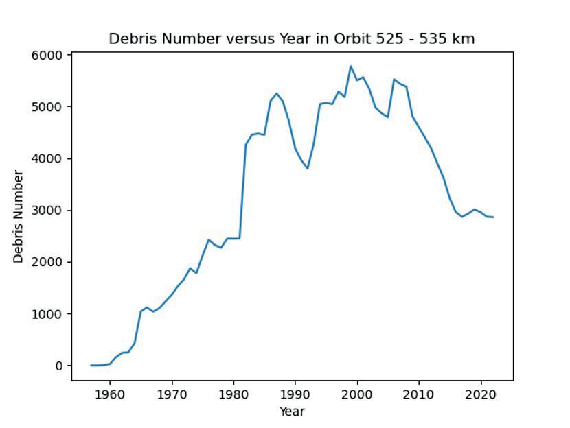
tion method can be used to process these data and then fit a curve consistent with the trend of growth to predict the number of debris in the future. However, an interpolation method might not always be precise to predict the number, especially after an extended period, as prediction numbers are based on logarithms. Instead, this paper predicts the number of debris in LEO over the next 18 years using a polynomial fit of the available data. The fitting curve with a polynomial degree of 3 is as Fig (4).
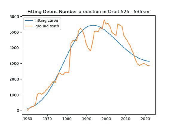
To obtain the fitted curve, the number of debris in the first three years since 1957 is not considered as the data affect the gradient of the fitted curve as the gradient of the fitting curved will be much steeper, which exaggerates the number for the predicted debris number. From the adjusted fitting curve, the number of debris in LEO in the next 18 years is predicted as Fig (5), where the predicted debris number at the region increased from 3,158 in 2022 to 6,287 in 2040.
Figure 3. Number of debris in altitude of 525-535km since 1957
47 Physics and Technology
Figure 4. The fitting curve of debris number against years since 1960
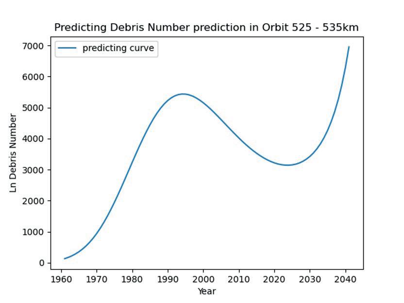
2.3 SpaceX Satellites Distribution
The number of Starlink satellites that can be launched is 12,000, and Starlink has applied to launch 30,000 more in the future. This set an upper bound for the number of Starlink satellites in LEO. There are over 3,300 Starlink satellites in LEO, including those just launched. The majority of Starlink satellites orbit between latitudes 53 to -53 degrees, with only about 3% of satellites crossing over the latitude between 53 to -53 degrees [11]. It is assumed that the Starlink satellite does not have an autonomous collision avoidance system under the model in this paper.
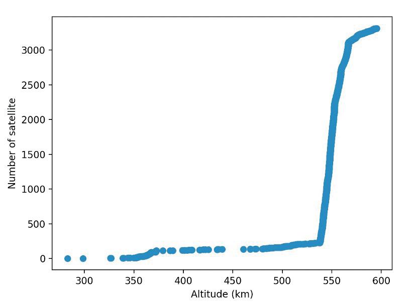 Figure 5. Predict number of debris from 1960 to 2040
Fig (6) shows that 89.7% of current Starlink satellites were at an altitude between 530 km to 600 km, where this research mainly focuses.
Figure 5. Predict number of debris from 1960 to 2040
Fig (6) shows that 89.7% of current Starlink satellites were at an altitude between 530 km to 600 km, where this research mainly focuses.
48 Scientific Harrovian 2022
Figure 6. Distribution for the altitude of Starlink satellite
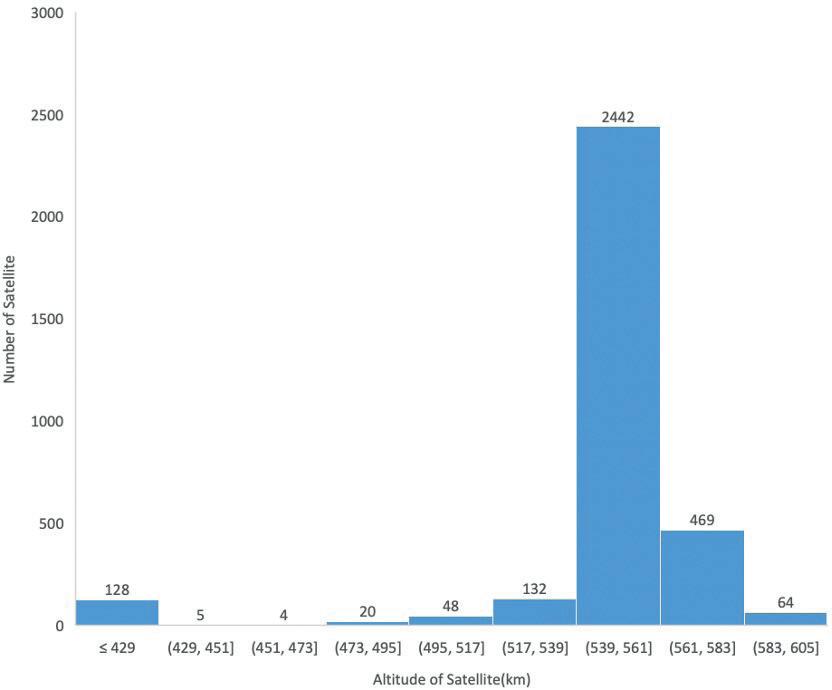
From fig (7), around 3,100 Starlink satellites lie between 500 - 600 km altitude, and 2,442 Starlink satellites were distributed at an altitude between 539 km to 561 km. An initial modal can be constructed on the attitude between 539-561 km, as Starlink satellites operate most densely in this altitude region.
Since the distribution and orbits of the satellites are relatively fixed, the density formula can be used to estimate the distribution density of Starlink satellites at a given altitude and derive the number of satellites present in the set height. In the formula, Nsatellite is the number of satellites in the selected region, REarth is the radius of Earth, Hhigh is the altitude of higher of the selected region, and Hlow is the altitude of lower of the selected region.
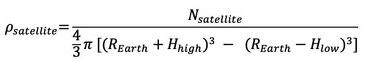
2.4 Dimension and Period of Satellite
All the current Starlink satellites in the LEO are generation 1 (Gen 1). Dimension of Gen 1 Starlink satellite has a length of approximately 2.8 meters, width of 1.4 meters, and height of roughly 0.2m [12]. The surface area of the Space X satellite solar panel is 23.657 (From the DISCOB web); this gives the solar length of the panel to 8.45m and the longest side of the satellite to be 8.65m. Approximation for the collision distance can be considered by using the dimension of the satellite.
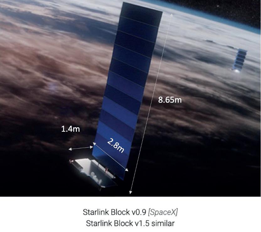
According to the dataset from 3,269 Starlink satellites, the average orbit period for a functional satellite is 95.06 minutes. This can be used to calculate the time length for a collision to occur after the construction of the model.
Figure 7. Bar graph for distribution of Starlink Satellite altitude
49 Physics and Technology
Figure 8. Gen 1 Starlink satellite (Picture reformated from [12])
Probability model based on Monte-Carlo Method
3.1 System Model
This paper proposes a model that utilizes the Monte Carlo method for practical application in the LEO space, which includes the Starlink satellite. The study focuses on a specific altitude within the LEO space by dividing LEO space into smaller sections of independent space for investigation purposes. The model is designed to maintain a constant satellite height during steady-state operation, disregarding physical parameters such as angular momentum and trajectory. To simulate the satellite coordinates in space, the satellite is approximated as a sphere with a fixed radius, and the sphere’s center point is used as the polar coordinates to determine the position of satellites relative to the independent space. Similarly, the space junk is approximated as a sphere with a uniform radius, and the center polar coordinates of the sphere are utilized to determine the position of the satellite with respect to the independent space. The number of debris and satellite in the set region is determined by debris and satellite density corresponding to the investigating region.
3.2 Application of the Monte-Carlo Method
With the density of the satellite and debris, an initial model can be built. The first step of the modal is to select the altitude interval to be investigated. Let the center of Earth as the center; a hollow sphere is formed, as Fig (9) shows.
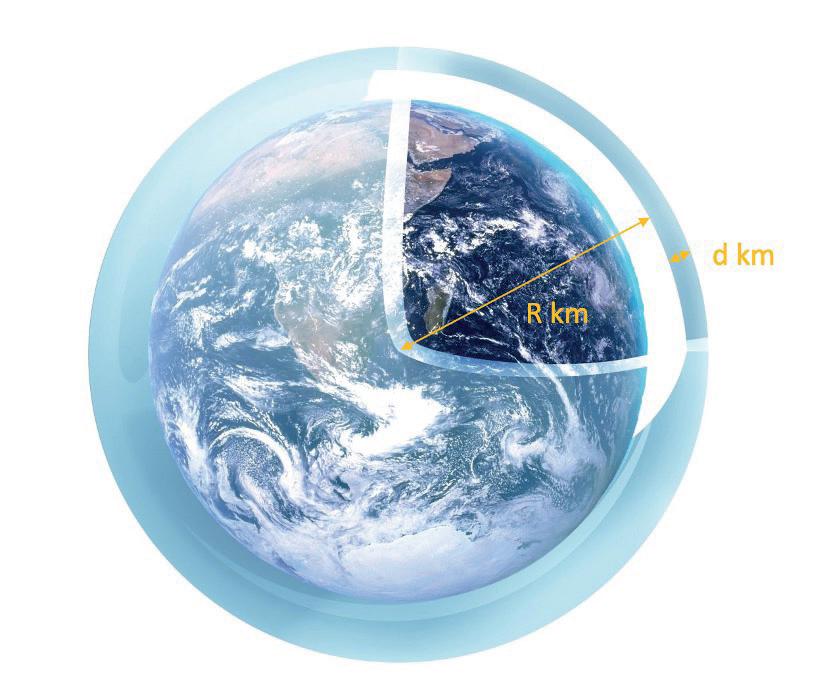
The thickness of the ‘sphere shell’ is the selected altitude interval where the difference in the altitude of the ‘sphere shell’ is defined by ‘d’. The distance from the center of the Earth to the lower bound of the ‘sphere shell’ is defined by ‘R,’ and the total volume of the ‘sphere shell’ is the volume of the selected altitude interval where Fig (10) shown.
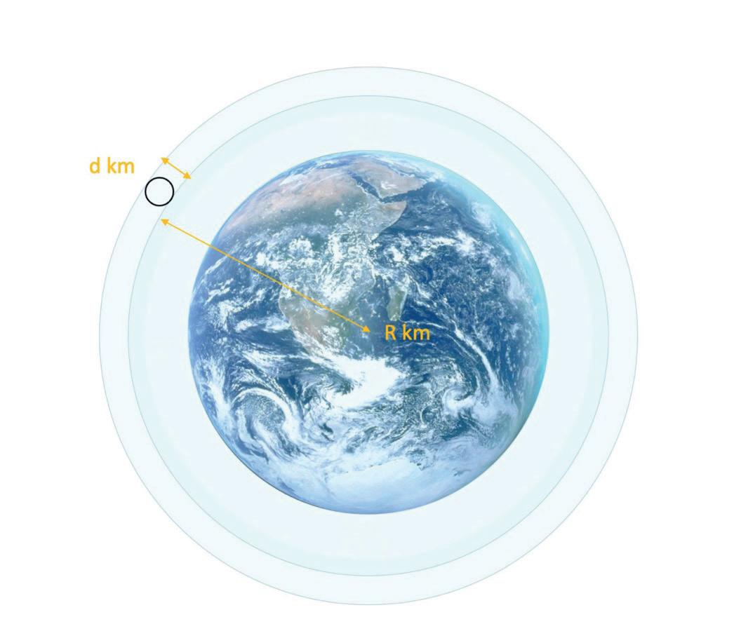 Figure 9. Starlink orbit shell around Earth
Figure 10. Cross-sectional view of the orbit
Figure 9. Starlink orbit shell around Earth
Figure 10. Cross-sectional view of the orbit
50 Scientific Harrovian 2022
To simplify the model, a circle is constructed inside the shell where the diameter of the circle is ‘d’. Extend the circle around the center of the shell to obtain a ‘doughnut’ shape space. This doughnut-shaped space, when stretched out, can form a cylinder space as shown in Fig (11). The height of the cylinder form is the circumference of the ‘doughnut’ shape. That is also the circumference of the sphere form with a radius from the center of the Earth to the upper bound of the ‘sphere shell.’ To determine the volume of the cylinder space form, the equation is as follows:
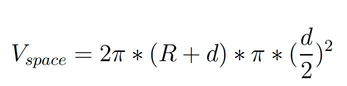
Where R is the distance from the center of Earth to the lower boundary of the ‘sphere shell,’ and d is the diameter of the cylinder form. Estimate number of Starlink satellites and debris in selected space is given as:
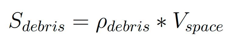
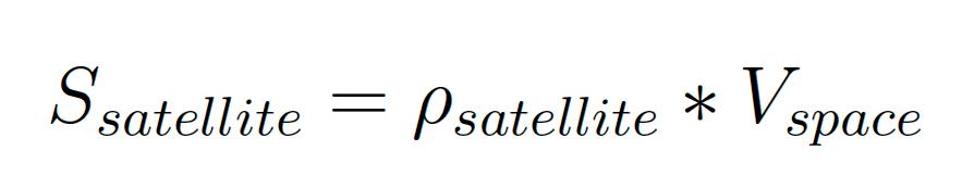
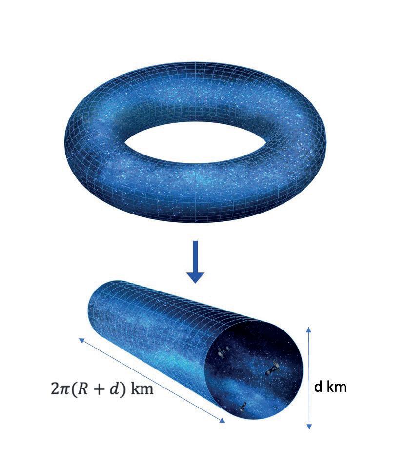
The model assumes that the satellite’s orbital altitude does not change during the operation cycle, so a single satellite moves in a straight line in the cylindrical space of the model. For any debris, the model assumes it is a stationary sphere in the cylinder. A circle is drawn with a radius defined as ‘safety distance’ with the center point of the satellite as the center of the circle. A smaller cylinder, defined as a ‘safety zone’, is obtained after the satellite moves from one side of the cylinder to the other side. The model defines whether the satellite collides with the debris as whether the sphere represented by the debris is in the safety
51 Physics and Technology
Figure 11. Uniform cylinder space for cross-sectional direction
zone formed by the movement of a satellite in cylindrical space. If the sphere represented by the debris is in the safety zone, the model determines that the satellite and the debris will collide and count number +1. After the previous settings, further simplification can be done, which is converting the model from 3d to a more concise 2d model. As the orbit of the satellite in the cylindrical space is known to be a straight line, the determination of whether the debris collides is made by observing whether the sphere represented by the debris appears in the safety zone formed by the satellite. The cylinder can then be compressed, and as the thickness of the cylinder space approaches zero, a circle of diameter d km, as shown in Fig (12), is formed.
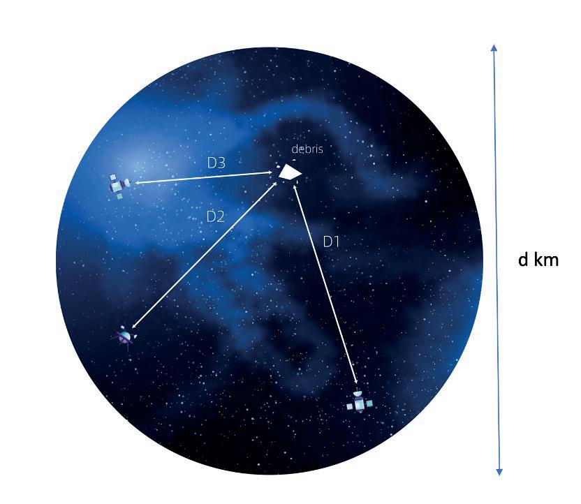
The model’s determination of whether there is a collision is more straightforward under 2d. As shown in Fig. (12), it is assumed that three satellites are on a circle with a diameter of d km, and their distance from the debris randomly appearing on the circle is D1, D2, and D3, respectively. When the distance between any D1, D2, or D3 is less than the specified safe distance, the model determines a collision. Polar coordinates determine the position of the satellite and random debris on the circle. At the start of each orbit, a random polar coordinate is given to the satellites and debris, and the coordinate is changed by random functions after each satellite completes one full orbit of motion. Since the selected cylindrical space is random in the ‘spherical shells,’ the position of the satellite and debris change after each orbit period. Although the position of the satellite and debris is changed randomly, it must be within the space of the circle formed by the model, as Fig (12) shows, and not beyond it, as this will cause changes in the distribution density of the debris and satellite.
Simulation and Result Analysis
4.1 Simulation Setup
Considering that the radius of the sphere formed by the satellite and the sphere formed by the random debris is close to 20 meters, and the relative velocity between debris and Starlink satellite in LEO is approximately 10 to 15 km (about 9.32 miles) per second [13]. In this model, 100 m is used as the standard safety distance to define whether there is a collision or not to reduce the uncertainty in the model due to simplification.
4.2 First Scenario: Constant debris numbers
The first scenario sets a constant debris number for the model and the satellite. During the repeat of the experiment, the number of debris in the model does not change as time changes. The number of debris in LEO to estimate the corresponding debris density was using the data in 2022, which is 3042. The number of satellites that are used to estimate satellite density is 2442. It is assumed that the distribution of satellites and debris is totally random in the given region. This model selected the altitude where Starlink satellites distribute most densely for an experiment. According to the altitude distribution of Starlink satellites in 52 Scientific Harrovian 2022
Figure 12. Simplification of 3d cylinder space to 2d
LEO, choose the region (539 km to 561 km). The density of Starlink satellites at this altitude is 1.840*10-7 Satellite/km3 . Depending on the model setup, a cylinder with a radius of 11 kilometers and a length of 43,486 kilometers will be formed. Based on the modal, in the attitude of 531km to 561km, each ‘cylinder’ contain 2.813 Starlink satellites and 3.493 debris. In the simplified model, three circles with a radius of 10 meters circle are placed randomly in a radius of 11 kilometers to represent the respective positions of the 2 satellites and an additional 0.813 probability of having an extra satellite appear. Meanwhile, there is 3 debris with an additional 0.493 probability that a circle with a radius of 0.1m be randomly placed on the circle to represent the position of random debris. The probability of collision is defined as the average number of orbits it takes for one collision to occur. By counting the average collision counts that occur during the 100000 periods of the orbit, the probability of collision can be determined from the following equation, where Pcollision is collision probability and Ncollision is the average collision count over 100000 periods.
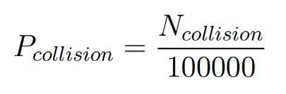
4.3 Second Scenario: Consider the increment of Satellite and Debris number
The second scenario considers the increment of satellite and debris as time changes. This model focuses on the altitude between where it is the region the Starlink satellite will be most densely distributed in the future. The number of debris in LEO to estimate the corresponding debris density is using the data from the predicted number of debris from 2022 to 2040, which is 3,158 to 6,287. The number of satellites that are used to estimate satellite density is 10,080. It is assumed that the distribution of satellites and debris is even in the given region. This model selected the altitude where Starlink satellites gather most densely for the experiment. With respect to the news, we chose the region (525km to 535km); the density of Starlink satellites at this altitude is 1.681*10-7 satellite/km3 . Depending on the model setup, a cylinder with a radius of 5 kilometers and a length of 43,360 kilometers is formed. Considering increased numbers of debris with the time change, the 100,000 orbit period of Starlink satellite is approximately 18 years. Taking the average for the predicted numbers of debris since 2022, the average estimated debris number is 3949.178. This led to the debris density in the selected region to be. The cylinder form contains 5.299 Starlink satellites and 2.076 debris. In the simplified model, 5 circles with a radius of 10 meters circle are placed randomly in a radius of 5 kilometers to represent the respective positions of the 5 satellites with an additional probability of 0.299 to have an extra satellite appear. Meanwhile, there is 2 debris with an additional probability of 0.076 that a circle with a radius of 0.1m be randomly placed on the circle to represent the position of random debris. The probability of collision can be determined by taking the average number of collisions that occur during the 100,000 periods of the orbit.
Result
With relevance to the model in the first scenario where the safety distance is 100m, the average count for the number of collisions is 84.84 per 100,000 periods. Under the definition of collision probability set in the scenario, the probability of collision is 8.484*10-4 Fig (13) shows how the collision count predicts change as safety distance varies in the range from 50 to 500 meters. From Fig (13), a non-linear growth is observed when the safety distance increase from 50m to 500m with the same experimental setup in scenario one. With the safety distance of 50m, average collision counts are 21.75, which increased to 2140.37 when the safety distance is 500m. The corresponding collision probability increased from 2.174*10-4 to 2.140*10-2.
53 Physics and Technology
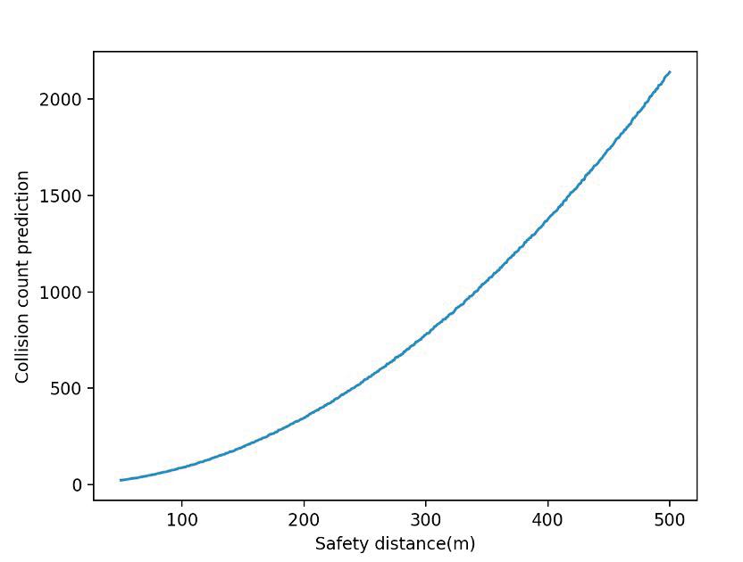
For the model in the second scenario, where the safety distance is 100m, the average count for the number of collisions is 452.99 per 100,000 periods. Under the definition of collision probability set in the scenario, the probability of collision is 4.530*10-3. Fig (14) shows how the predicted collision counts change as safety distance varies in the range from 50 to 500 meters. From Fig (14), a non-linear growth is observed when the safety distance increase from 50m to 500m with the same experimental setup in scenario one. With the safety distance of 50m, average collision counts are 117.33, and this increased to 11157.32 when the safety distance is 500m. The corresponding collision probability increased from 1.173*10-3 to 1.116*10-1.
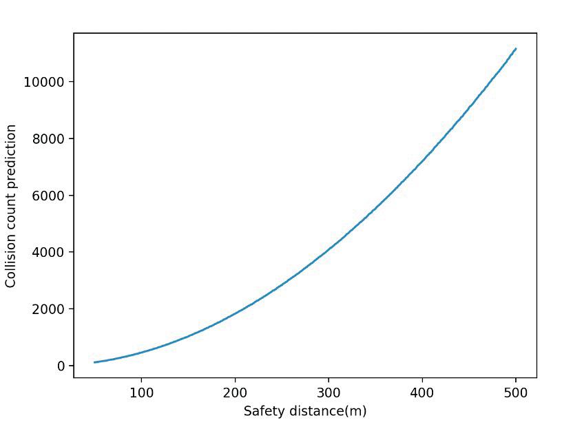
When comparing the data from both scenarios. In the same safe distance, the growth rate of predicted collision counts in scenario 2 is significantly higher than that in scenario 1, as shown in Fig (15). Within the set safe distance of 50 to 500 meters, the average number of collisions in scenario 2 is almost five times that in scenario 1 at the same safety distance. Considering the uncertainty brought by the estimation and approximation of the model, an uncertainty test is also applied to the prediction of the collision counts. By using standard error, a graph of standard error against safety distance set from 50 to 500 m is plotted as Fig (15) shows. For the first scenario, at the safety distance of 50m, the standard error is 0.4723, where the error increases to 4.3024 when the safety distance is set at 500m. For the second scenario, the standard error at 50m is 0.9650 and increases to 11.0737 when the safety distance is set at 500m. Although the standard error increased linearly along the increasing safety distance for both scenarios, this does not affect the non-linear growth for the collision counts when the safety distance increases.
Figure 13. Collision count prediction against safety distance for the first scenario
54 Scientific Harrovian 2022
Figure 14. Average collision counts against safety distance for the second scenario
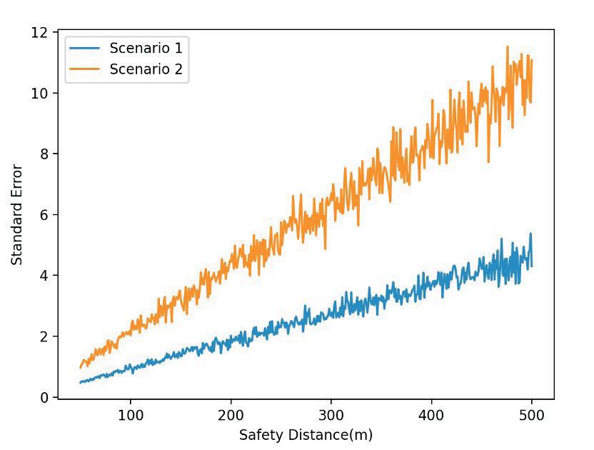
Conclusion
This paper estimates the current probability of debris colliding with Starlink satellites at the altitude where the satellites are most densely clustered. The average collision probability of Starlink satellites at most densely clustered in the next 18 years is also given. The model in this paper assumes that the Starlink satellite does not have an autonomous collision avoidance system and ignores the physical quantities such as velocity and time affecting the satellite when calculating the collision probability. The Monte Carlo method is used to estimate the probability of collision after simplification of the initial model. Considering the error caused by model simplification and approximation, the standard safety distance of the model is set to 100 meters. At altitudes of 539 to 561 kilometers, the Starlink satellite had a collision probability of 8.484*10-4 in 2022. The estimated number of collisions increases nonlinearly with the increase in safe distance. Over the next 18 years, the Starlink satellite collided with debris at an altitude of 525 to 535 kilometers with an average probability of 4.530*10-3 if the orbit is filled with the estimated number of satellites. Also, the estimated number of collisions also presents a nonlinear increase with the increasing safety distance. In the analysis of the predicted collision counts, the standard error of the predicted collision counts increases with the increase of the safety distance, but the error range does not affect the nonlinear increase of the predicted collision counts. It is concluded that the probability of collision rises significantly with the increased safety distance set, and the standard error of the experiment rises linearly along the increasing average collision count. Therefore, and it is concluded that in the long run, the increase of collision probability with time has posed a significant risk to the safety of Starlink satellites. Therefore, it is necessary to continuously develop and improve the debris detection and collision avoidance system in the future.
The advantage of the model in this paper is that it uses a relatively simple method and model to give a conservative collision probability. First, by simplifying approximate to a certain extent, a 3d collision problem is converted into 2d, which makes the model more visualization. Second, the computation can also be greatly reduced by ignoring the physical quantity that could affect collision probability, as well as not using calculus and matrices. This method can be used to rapidly estimate the collision probability of satellites and debris within a specific chosen region. Finally, this method is suitable for the non-expert. Due to the model’s simplicity, it can provide a more intuitive understanding of the collision between the satellite and debris of the public. The experiments described in the paper can also be performed on a commercial computer, which means it is available to the public.
Although the model can easily and quickly predict the collision probability, adjustment to improve the accuracy of prediction is also vital. There are several methods that can increase the accuracy of the model.
Figure 15. Standard error with safety distance increase for both scenarios
55 Physics and Technology
First, the model in our paper uses the average density of Starlink satellites to estimate Starlink satellites in each cylindrical space. However, because most Starlink satellites are not distributed in high and low latitudes, the density of the satellites will be different from that calculated in the paper when the ‘doughnut’ model space is used to simplify, resulting in a different number of satellites in each ‘doughnut’ space.
Then, if the high and low latitude spaces are removed, the original model is also no longer a spherical shell around the Earth, resulting in a different volume of each doughnut space formed in the model. Therefore, considering the number density distribution function is necessary to improve the experimental accuracy. But at the same time, a lot of integration is required, and more accurate mathematical models to approximate the geometrical shape of the shell.
Second, the model’s definition of the collision range of the satellite and debris could be more rigorous. The collision range of this model is determined to be a collision when any debris appears in the safety zone formed on the orbit of the satellite. The prerequisite is to ignore the fact that the coordinates of the satellite change over time in cylindrical space. If the influence of time change on the real-time coordinates of the satellite is considered, it will be more accurate to use the matrix to represent the coordinate of the satellite in the cylinder space at any time, and the distance between random debris and the satellite at a particular time can be calculated more intuitively.
Finally, in future research, in order to predict the collision probability more reasonably and accurately, considering the collision avoidance maneuvers performed by the satellites that mitigate the probability of collision will be necessary. Take the Starlink satellite as an example. In the report from SpaceX, during the period from June 1, 2022 – November 30, 2022, Starlink satellite has performed 13,612 propulsive maneuvers, which is approximately 12 maneuvers per satellite [14]. It is noticed that for Starlink satellite, if collision probability reaches above 1*10-5, then maneuvers must be taken. Such maneuvering would significantly reduce the likelihood of collision and should be considered in future studies.
Supplemental information
Link to the online code database: https://github.com/CheukHonLi/Starlink_Satellite_Probability_Collision_Model
References
[1] https://www.ucsusa.org/resources/satellite-database
[2] https://aviation-edge.com/spacex-satellite-tracker-api/
[3] https://www.space.com/spacex-starlink-satellites.html
[4] https://www.esa.int/Space_Safety/Space_Debris/Space_debris_by_the_numbers
[5]https://www.esa.int/ESA_Multimedia/Images/2021/03/The_history_of_space_debris_creation#.ZFUTdpQ0wVQ.link
[6] https://www.space-track.org/#catalo
[7] Kelso, T. S. “Analysis of the Iridium 33-Cosmos 2251 collision.” Advances in the Astronautical Sciences 135.2 (2009): 1099-1112.
[8] Stevenson, Matthew, et al. “Identifying the statistically-most-concerning conjunctions in leo.” 2021 Advanced Maui Optical and Space Surveillance Technologies Conference (AMOS), Maui, Hawaii. 2021.
[9] Haicheng Tao, Xueke Che, Qinyu Zhu, XinHong Li, “Satellite In-Orbit Secondary Collision Risk Assessment”, International Journal of Aerospace Engineering, vol. 2022, Article ID 6358188, 18 pages, 2022. https://doi.org/10.1155/2022/6358188
[10] Patera, Russell P. “General method for calculating satellite collision probability.” Journal of Guidance, Control, and Dynamics 24.4 (2001): 716-722.
[11] https://satellitemap.space/?constellation=starlink0#
[12] https://space.skyrocket.de/doc_sdat/starlink-v1-5.htm
[13] https://www.nasa.gov/news/debris_faq.html
[14] https://planet4589.org/astro/starsim/docs/Star2212.pdf
56 Scientific Harrovian 2022
57 Physics and Technology

CHEMISTRY BIOLOGY and
58 Scientific Harrovian 2022

59 Chemistry and Biology

Do you want to LOOK YOUNG?
by Bernice Ho
60 Scientific Harrovian 2022
How would you feel if you had the skin of a 20-year-old when in reality you are 50? Many would love to attain this level of collagen in their skin. Not long ago, scientists in Cambridge successfully 1rejuvenated a 53-year-old woman’s skin cells to the equivalent of a 23-year-old’s using cell reprogramming.
This newfound success could not only contribute to the cosmetic field, but also help future generations look younger.
This article will explain to you the start of cellular reprogramming, the current findings and the future of how this one of a kind technology could benefit everyone’s health and happiness.
The Origin of Cellular Reprogramming
Cellular reprogramming is the process of converting a mature, specialised cell into an embryonic-like stem cell. The concept of rejuvenation and cellular reprogramming was first proposed by John Gurdon in the 1960s. He removed the nucleus of a fertilised egg cell from a frog and replaced it with the nucleus of a cell taken from a tadpole’s intestine, where the modified egg cell grew into a new frog.
In 1997, a team of scientists and researchers led by Professor Sir Ian Wilmut at the Roslin Institute in Edinburgh cloned an adult sheep called Dolly. This research breakthrough grabbed headlines around the world. Wilmut’s team tried to develop a better method to produce genetically modified livestock. 2 By turning an adult mammary gland cell taken from a sheep into an embryo, Dolly was created. 3
She was the first mammal to be cloned from an adult cell. Dolly’s birth proved that specialised cells could be used to create an exact copy of the animal they came from. With this knowledge, it proved that turning a differentiated cell back into any kind of cell could open up many opportunities, especially in biology and medicine, for example, treating diseases like spinal cord injury.
Gene regulatory networks and tissue morphogenetic events drive the emergence of shape and function, allowing scientists to mimic and manipulate human embryos. This also means that we could potentially turn an old cell into a younger cell.
In 2006, a new method, Induced Pluripotent Stem cells (iPS), was discovered by Shinya Yamanaka. iPS cells have the potential to develop into every type of cell in the body and are valuable tools for disease modelling, drug screening, and cell therapy. 4iPS cells also provide further opportunities for discovery in life science such as creating great potential in regenerative medicine.
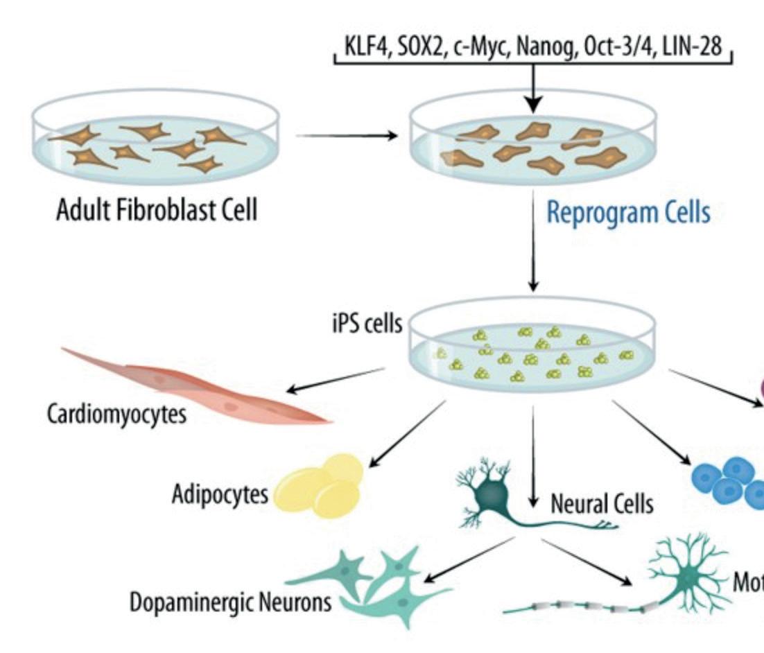

Introduction
61 Chemistry and Biology
Figure 1: Directed Differentiation of iPS Cells
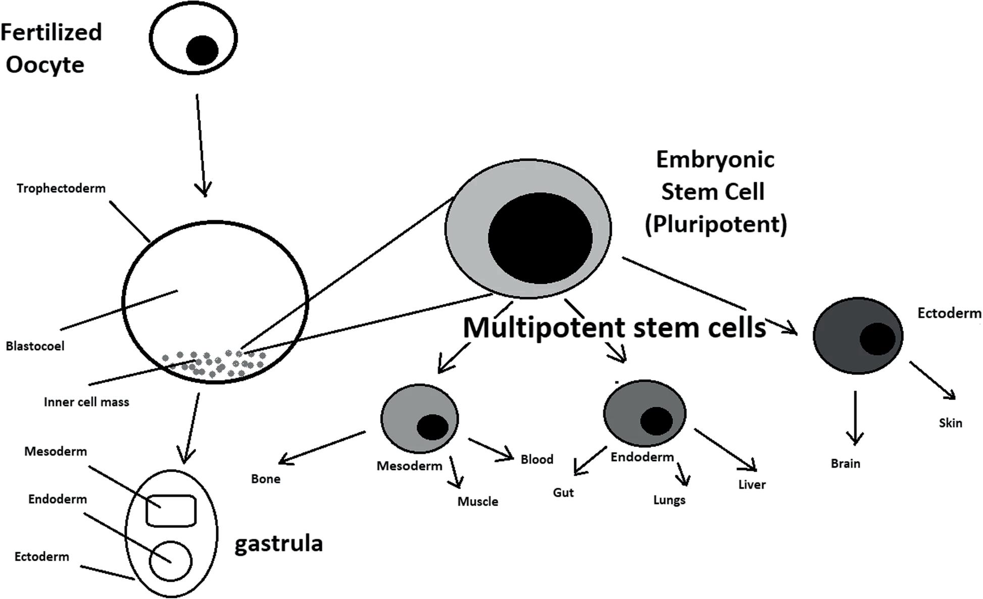
The aim of cell reprogramming is to convert 5stem cells to desired cell types. The most direct way of differentiating 6stem cells is to mimic the development of an inner cell mass during gastrulation. During gastrulation, pluripotent stem cells differentiate into ectodermal, mesodermal, or endodermal progenitors. Mall molecules or growth factors induce the conversion of stem cells into appropriate progenitor cells, which will later give rise to the desired cell type. But, of course, there are many other ways of differentiating a stem cell.
Turning Back the Ageing Clock
On 8th April 2022, scientists from the Babraham Institute, and a life sciences research institute in Cambridge, published an article on eLifesciences. The research team successfully 7rejuvenated a 50-year-old woman’s skin cells into behaving and looking as if they came from a 23-year-old in only 13 days.
Not only have they furthered the use of IPS cells, they have even developed the 8first ‘maturation phase transient reprogramming’ (MPTR) method, where reprogramming factors were expressed until a certain rejuvenation point followed by withdrawal of their induction. Cells’ fibroblast identity was found to be temporarily lost and then re-acquired during MPTR. This could be a result of epigenetic memory at enhancers and/or persistent expression of some fibroblast genes through using dermal fibroblasts from middle age donors. Amazingly,9 their method substantially rejuvenated multiple cellular attributes including the transcriptome (the array of mRNA transcripts produced), which was rejuvenated by around 30 years as measured by a novel transcriptome clock (Aging clock dissociates biological from chronological age).
However, for now, 10the MPTR technique will not be ready for use in clinics due to the potential increases in the risk of cancer brought on by genetic changes within the cells. Yet, scientists are confident that they can find a safer method to rejuvenate cells, and believe that they can apply the same technique to other tissues in the body. Ultimately, scientists are hoping to develop treatments for age-related diseases such as diabetes, heart diseases, and neurological disorders.
Figure 1 shows how iPS cells could differentiate into specialised cells like neural cells, Adipocytes and Cardiomyocytes etc.
Figure 2: Stem cell biology
62 Scientific Harrovian 2022
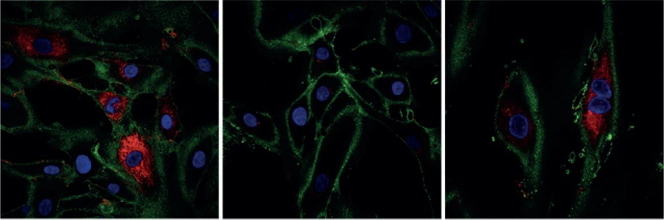
https://www.sciencealert.com/scientists-rewind-the-age-of-human-skin-cells-back-30-years
Potential applications of cell reprogramming
Cell reprogramming has the potential to become huge in the medical field as iPS cells play a vast role in developing restorative medicine. 11Serious medical conditions, like cancer, are caused by improper differentiation or cell division. Even though cell reprogramming isn’t vastly used to cure cancer, currently, several stem cell therapies are possible, among which are treatments for spinal cord injuries and heart failure12.
By furthering study in cell reprogramming, we may be able to find a cure for chronic diseases. One example is Alzheimer’s disease (AD), which is the most prevalent age-related dementia in the world. The underlying mechanisms of AD remain unclear. In recent years, upon the improvement of induced pluripotent stem cell technology and direct cell reprogramming technology, it has become possible to induce non-neuronal cells, such as fibroblasts or glial cells, directly into neuronal cells in vitro and in vivo. The induced neuronal cells are functional and can integrate into the local neural net. These incredible findings are encouraging and can provide a new clinical approach to treating AD.
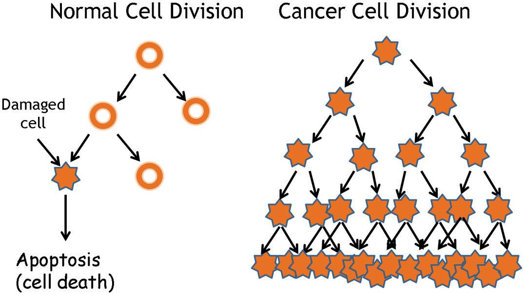
https://www.kindpng.com/imgv/hbwmixJ_picture-cancer-vs-normal-cell-division-hd-png/
Other than being able to use cell reprogramming in the medical field, 13cell reprogramming also gives a plausible answer for avoiding the use of human embryonic cells in experimental research and clinical medicine, which is ethically unacceptable, as obtaining these cells requires the destruction of human embryos. Cell reprogramming is considered a much better option than the use of embryonic stem cells.
Figure 2: Collagen production (in red) being restored in cells after reprogramming. (Fátima Santos, Babraham Institute)
Figure 3: Normal cell division vs cancer cell division
63 Chemistry and Biology
Cell reprogramming has led to many new findings, for example, cell rejuvenation of a 50-year’s old woman’s skin cells into a 23 year old’s skin in a very short period of time. Even though cell rejuvenation is just a very small part of cell reprogramming, there are many things to explore within this research field. Hopefully, in the near future, cell reprogramming will be able to be used in many different therapeutic areas to improve our healthy lifespan and save thousands of lives.
Bibliography
[1]“Rejuvenation of Woman’s Skin Could Tackle Diseases of Ageing.” BBC News, 8 Apr. 2022, www.bbc.com/news/science-environment-60991675.
[2]“Rejuvenation of Woman’s Skin Could Tackle Diseases of Ageing.” BBC News, 8 Apr. 2022, www.bbc.com/news/science-environment-60991675.
[3]Roslin Institute. “The Life of Dolly | Dolly the Sheep.” Ed.ac.uk, 2019, dolly.roslin.ed.ac.uk/facts/the-life-of-dolly/index.html.
[4]“Shinya.yamanaka@Gladstone.ucsf.edu.” Gladstone.org, gladstone.org/people/shinya-yamanaka#:~:text=In%202006%2C%20Shinya%20 Yamanaka%20discovered.
[5]Zakrzewski, Wojciech, et al. “Stem Cells: Past, Present, and Future.” Stem Cell Research & Therapy, vol. 10, no. 1, 26 Feb. 2019, stemcellres. biomedcentral.com/articles/10.1186/s13287-019-1165-5, 10.1186/s13287-019-1165-5.
[6]Zakrzewski, Wojciech, et al. “Stem Cells: Past, Present, and Future.” Stem Cell Research & Therapy, vol. 10, no. 1, 26 Feb. 2019, stemcellres. biomedcentral.com/articles/10.1186/s13287-019-1165-5, 10.1186/s13287-019-1165-5.
[7]“Rejuvenation of Woman’s Skin Could Tackle Diseases of Ageing.” BBC News, 8 Apr. 2022, www.bbc.com/news/science-environment-60991675.
[8]Gill, Diljeet, et al. “Multi-Omic Rejuvenation of Human Cells by Maturation Phase Transient Reprogramming.” ELife, vol. 11, 8 Apr. 2022, 10.7554/elife.71624. Accessed 13 Apr. 2022.
[9]Gill, Diljeet, et al. “Multi-Omic Rejuvenation of Human Cells by Maturation Phase Transient Reprogramming.” ELife, vol. 11, 8 Apr. 2022, 10.7554/elife.71624. Accessed 13 Apr. 2022.
[10]“Rejuvenation of Woman’s Skin Could Tackle Diseases of Ageing.” BBC News, 8 Apr. 2022, www.bbc.com/news/science-environment-60991675.
[11]Zakrzewski, Wojciech, et al. “Stem Cells: Past, Present, and Future.” Stem Cell Research & Therapy, vol. 10, no. 1, 26 Feb. 2019, stemcellres. biomedcentral.com/articles/10.1186/s13287-019-1165-5, 10.1186/s13287-019-1165-5.
[12]Menasché, Philippe, et al. “Human Embryonic Stem Cell-Derived Cardiac Progenitors for Severe Heart Failure Treatment: First Clinical Case Report: Figure 1.” European Heart Journal, vol. 36, no. 30, 19 May 2015, pp. 2011–2017, academic.oup.com/eurheartj/article/36/30/2011/2398140, 10.1093/eurheartj/ehv189.
[13]Aznar Lucea, Justo, and Miriam Martínez. “[Ethical Reflections on Cell Reprogramming].” Cuadernos de Bioetica: Revista Oficial de La Asociacion Espanola de Bioetica Y Etica Medica, vol. 23, no. 78, 2012, pp. 287–299, pubmed.ncbi.nlm.nih.gov/23130744/. Accessed 29 Nov. 2022.
Conclusion
64 Scientific Harrovian 2022
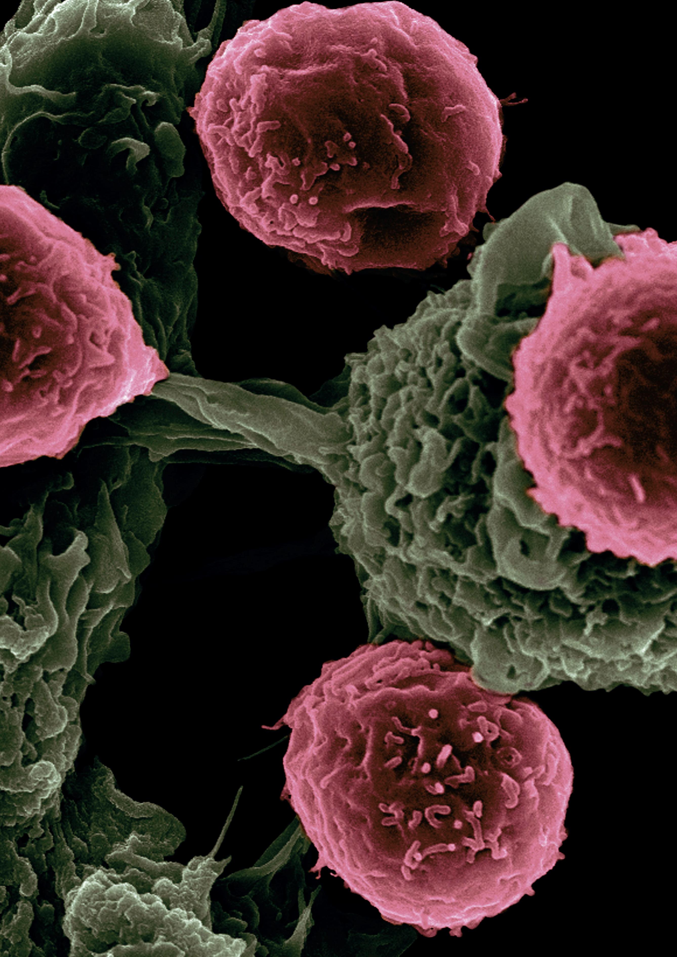
New Treatments for CANCER ?
by Audrey Lai
65 Chemistry and Biology
Nearly 10 million people die from cancer every year, making it the second most common cause of death in the world.1 Chemotherapy, radiation therapy, and surgery have been the traditional treatments for cancer since the 20th century and are still commonly used today. However, as many already know, the effectiveness of these traditional cancer treatments is often limited. Thanks to the quick development of technologies and medical discoveries, there are currently new, promising methods for treating cancer which includes immunotherapy, a modern medical innovation. In this article, we will explore what cancer is, how traditional cancer treatments work, and why we need immunotherapy, such as monoclonal antibodies and CAR T-cell therapy. First of all, what is cancer?
Cancer
Cancer is a non-communicable disease in which body cells grow and divide uncontrollably, spreading to other parts of the body via the bloodstream. Human body cells undergo mitosis, also known as cell division, in order to grow and repair old, damaged cells. A mutation in the gene that regulates a cell’s functions and division results in cancer. Genes involved in regular cell growth can become oncogenes or healthy tumour suppressor genes can become inactive as a result of a change in the DNA. Consequently, this mutation will cause uncontrolled cell growth. Gene mutations can result from random errors during cell division, damage to DNA caused by carcinogens such as tobacco, or inheritance from one’s parents. Cancer cells that have detached from their primary tumour and travelled through the bloodstream to form secondary tumours in other parts of the body are said to have metastasized.
Traditional Treatments
Surgery, chemotherapy, and radiation have always been popular options in treating cancer, yet the chances of a patient with cancer dying of it are similar to those of 50 years ago.2 First, chemotherapy works by using chemicals to shrink tumours, destroy or kill cancer cells, and help other treatments work better. However, the main drawback of chemotherapy is that it simultaneously kills cancer and healthy cells that grow and divide quickly, such as hair, skin, blood and intestinal cells. As a result, it leads to potential side effects like hair loss, nausea, fatigue, and infections. Second, radiation therapy works by using radiation to shrink tumours and slow the growth of cancer cells by damaging their DNA. It is a local treatment and can be given externally or internally. The disadvantages include damaging surrounding healthy cells, leading to side effects and patients also have a lifetime dose limit to the amount of radiation that can be received by the body. Third, surgery is another option for removing tumours grown from cancer cells. A limitation of surgery is that blood cancer or metastatic cancer that has spread cannot be treated with surgery. This makes surgery only available for removing solid, local tumours in the body. Therefore, targeted therapy like immunotherapy is designed to treat targeted cancer cells.
Immunotherapy
The immune system helps detect and destroy any foreign or abnormal cells in our body including cancer cells. Immunotherapy is a type of cancer treatment, which aims to encourage the patient’s immune system to target cancer cells and destroy them. The treatments are designed to target a specific antigen on a specific type of cancer cell while sparing healthy body cells. Immunotherapy includes cancer vaccines, adoptive cell therapy (CAR T-cell therapy), checkpoint inhibitors and monoclonal antibodies. Over 100 different types of cancer exist today, which is a significant number. 3Therefore, since each type of cancer can be significantly distinct, a more targeted and focused approach is needed.
Introduction
66 Scientific Harrovian 2022
Monoclonal Antibodies
One of the popular methods of immunotherapy is monoclonal antibodies. Did you know that the EU and the US have approved around 100 therapeutics monoclonal antibodies for treating both cancer and non-cancer diseases? 4Monoclonal antibodies are becoming more and more common in the field of medicine due to their ability to bind to a specific protein or antigen on cell membranes. Other applications of monoclonal antibodies include the delivery of medications to cancer cells, the attachment of fluorescent substances to detect specific cells, pregnancy testings, and the diagnosis of numerous diseases such as HIV.
How are monoclonal antibodies produced?5
1. A type of antigen on the cancer cell is injected into mice and their B-cell lymphocytes are stimulated to produce the antibodies for the specific antigen.
2. The mice undergo blood screening for the antibody production.
3. The splenocytes which produce the B-lymphocytes are removed.
4. The splenocytes are fused with myeloma cells, which can divide unlimitedly, forming hybridoma cells.
5. The hybridoma cells divide and produce many clones specific to cancer’s antigen.
6. The clones are screened and selected, then purified. As a result many clones of antibodies are made.
How do monoclonal antibodies work in the body?
1. Monoclonal antibodies are injected into the patient’s bloodstream.
2. They will locate proteins called antigens on cancer cells.
3. Since the monoclonal antibodies are specific to the antigen on cancer cells they will bind and form an antibody-antigen complex.
4. The antibody will then signal other immune cells.
5. The immune cells will arrive and help destroy cancer cells.
Monoclonal antibodies treatment in cancer:
In recent years, monoclonal antibodies have become an option for treating cancers. An example is rituximab (Rituxan), a monoclonal antibody targeting leukaemia and B-Cell non-Hodgkin lymphoma (NHL). According to a study, patients with lymphomas who received rituximab and chemotherapy had a better survival rate than those receiving chemotherapy.6
Car T-Cell Therapy
Did you know since 2017, six CAR T-cell therapies have been approved by the FDA for the treatment of blood cancers? 7Acute lymphocytic leukaemia has a 40% 5-year survival rate for people aged 20 and older.8 However, blood cancer is now incredibly manageable due to decades of research and the introduction of innovative treatments like CAR T-Cell therapy.
How are CAR T-cells made:
1. Blood is drawn from a patient which moves through a blood separator to collect the T-cells, the remaining blood components will return to the blood.
2. The T-cells are genetically engineered by editing their gene to produce a protein on their surface (chimeric antigen receptors) which bind to specific proteins or antigens on the cancer cells.
3. Genetically modified T-cells are grown until there are millions of them and then they are collected.
How CAR T-cells work in the body:
1. After producing the CAR T-cells, they are reinfused back into the patient’s blood.
2. In the body, the CAR T-cells will bind to proteins on the cancer cells.
3. This will signal the immune system to destroy them.
67 Chemistry and Biology
CAR T-cell therapy treatment in cancer:
tisagenlecleucel is a CAR T-cell medication to treat B-cell acute lymphoblastic leukaemia. In 2021, a patient in Hong Kong was successfully treated with tisagenlecleucel (Kymriah). 9After relapsing twice from chemotherapy and haematopoietic stem cell transplantation, he was recommended CAR-T cell therapy. After the treatment, he recovered without experiencing any serious complications and results from bone marrow examinations show that no leukaemia cells could be found.
Modern VS Traditional
On one hand, immunotherapy seems to be a modern, better treatment due to its capability to target particular cancer cells while sparing healthy cells. This makes patients receiving targeted therapy experience fewer and less severe side effects compared to chemotherapy. However, there are a lot of drawbacks to the treatment. Decades of study, several pharmacological trials, and substantial financial resources are needed to develop a drug. Hence, immunotherapy is quite costly and out of reach for many individuals. For example, targeted therapy drugs like trastuzumab (Herceptin) for breast cancer costs $500,000 HKD per year and sorafenib (Nexavar) for liver cancer costs $150,000 HKD per month.10 This raises ethical issues because it is unfair that only wealthy people are able to access the latest advanced treatments for cancer. Conversely, there are also some highly effective traditional treatments even when they cannot specifically target cancer cells. In order to reduce costs and give all citizens an equal chance to obtain cancer treatment, a possibility is for the government to invest more in subsidising cancer treatment research. To conclude, both traditional and immunotherapy treatments for cancer could be used.
Bibliography
[1] WHO International Agency for Research on Cancer. “WHO International Agency for Research on Cancer.” “Cancer Today.” Published 2020. Global Cancer Observatory, https://gco.iarc.fr/today/online-analysis-pie?v=2020&mode=population&mode_population=income&population=900&populations=900&key=total&sex=0&cancer=39&type=1&statistic=5&prevalence=0&population_group=0&ages_ group%5B%5D=17&nb_items=7&group_cancer=1&include_nmsc=1&include_nmsc_other=1&half_pie=0&donut=0.
[2] Goodman, Amy, et al. “Why the ‘Slash-Poison-Burn’ Approach to Cancer Has Failed.” Truthout, Truthout, 23 Dec. 2019, https://truthout. org/video/why-the-slash-poison-burn-approach-to-cancer-has-failed/.
[3] “What Is Cancer?” National Cancer Institute, https://www.cancer.gov/about-cancer/understanding/what-is-cancer#:~:text=There%20 are%20more%20than%20100,cancer%20starts%20in%20the%20brain.
[4] “UpToDate.” Www.uptodate.com, www.uptodate.com/contents/overview-of-therapeutic-monoclonal-antibodies/print#:~:text=Since%20 1985%2C%20approximately%20100%20monoclonal.
[5] “Monoclonal Antibody Production.” Molecular Devices, https://www.moleculardevices.com/applications/monoclonal-antibody-production#gref.
[6] Schulz, Holger, et al. “Chemotherapy plus Rituximab versus Chemotherapy Alone for B-Cell Non-Hodgkin’s Lymphoma.” Cochrane Database of Systematic Reviews, vol. 2010, no. 1, 17 Oct. 2007, www.ncbi.nlm.nih.gov/pmc/articles/PMC9017066/, 10.1002/14651858.cd003805.pub2.
[7] “Car T Cells: Engineering Immune Cells to Treat Cancer.” National Cancer Institute, https://www.cancer.gov/about-cancer/treatment/research/car-t-cells#:~:text=Since%202017%2C%20six%20CAR%20T,%2C%20most%20recently%2C%20multiple%20myeloma.
[8] “Leukemia - Acute Lymphocytic - ALL - Statistics.” Cancer.net, 25 June 2012, www.cancer.net/cancer-types/leukemia-acute-lymphocytic-all/ statistics#:~:text=The%205%2Dyear%20survival%20rate%20for%20people%20age%2020%20and.
[9] “HKUMED Introduces Hong Kong’s First Car-T Cell Therapy for Blood Cancer Patients.” HKU Li Ka Shing Faculty of Medicine, 10 Feb. 2021, https://www.med.hku.hk/en/news/press/20210210-hk-first-car-t-cell-therapy-for-blood-cancer-patients.
[10] “Targeted Therapy Drug: Cancer Treatment, Cost & Funding in Hong Kong.” Www.cigna.com.hk, www.cigna.com.hk/en/smarthealth/medical/targeted-therapy-drug-cancer-treatment-cost-funding-in-hong-kong.
68 Scientific Harrovian 2022
Sustainable Life Through the ART BUILDING DESIGN of
by Kate Xiao

69 Chemistry and Biology
A brief summary of the principles behind carbon neutral buildings and how they will help us establish a cleaner way of living.
Assessing the environmental impact of buildings
In the contemporary world, buildings are responsible for more than 40% of energy usage and 33% of total greenhouse gas emissions in both developed and developing countries.1 According to the U.K Green Building Council, the construction sector uses more than 400 million tons of materials a year, many of which, such as aluminium, concrete, and steel, have an adverse impact on the environment through their high concentration of embodied carbon content (which refers to the greenhouse gas emissions arising from the manufacturing, transportation, installation, maintenance and disposal of a specific building material), with 9.8 million tons of CO2 produced by 76 million tons of finished concrete in the US.2
Fortunately, this data has not gone unnoticed. In the UK, after the launch of the Green Guide to Specification, Oxford Brookes University and the UK construction industry set out regulations to use certain materials in order to reduce the environmental impact, 230,000 construction projects have improved their environmental standing.2 In the US, the Environmental Protection Agency(EPA) also published a number of rules to reduce negative environmental effects (which includes areas such as soil stabilization, erosion and sediment controls, water contamination and waste management). The EPA also has a Greenhouse Gas Reporting Program (GHGRP), where relevant information is gathered every year from large GHG emission sources and reported to the public every October. This can help organisations and companies keep track of GHG emission states and alter the way of maintenance accordingly.
Before the construction of a building, a life cycle assessment (LCA) is carried out to determine the environmental impact it will bring during its lifetime, from designing to demolishment. A LCA is able to assess energy consumption and environmental impact through a scope of analysis with each type of building and fabrication method and each type of manufacturing or building material. With acknowledgement to different effects building materials and dimension choices will bring, designers can alter their scheme at an early stage and plan to embody the most impact reductions as possible.
What is a carbon neutral building?
The definition of a carbon neutral building is a building specifically designed to minimise greenhouse gases at all stages. As opposed to the commuter towns left from the Industrial Revolution and rapid urban development in the 21st century, a new trend in urban planning is led by urban projects that aim to reduce the negative impacts on the environment: The construction of sustainable buildings, carbon neutral buildings, eco-neighbourhoods, and more is highly commended on an international basis.
The principles behind carbon neutral buildings
If other buildings aim to reduce its environmental impact to the minimum, carbon neutral buildings aim to achieve net zero carbon emission. All the climate budget from materials manufacturing, transportation, construction process, and building in operation will be counteracted by investing in renewable energy, using timber as carbon storage, engaging in innovative and novel environmentally-friendly technology (examples are listed in case study below). The point is that over the years, net carbon emission remains zero, achieving carbon neutrality.
70 Scientific Harrovian 2022
Case study: Hong Kong
In Hong Kong, the first carbon neutral building—CIC-Zero Carbon Park—was built in 2012.3
Aside from serving as an exhibition, education and information center, CIC-ZCP acts as a test bed for state-of-the-art eco-building design. A native urban woodland in the middle of the compact, densely populated city, 47% of CIC-ZCP land area is covered in greenery, attracting birds and other animals to create a more biodiverse environment.3 The whole building relies on on-site renewable energy generated from photovoltaic panels and a tri-generation system using biofuel.
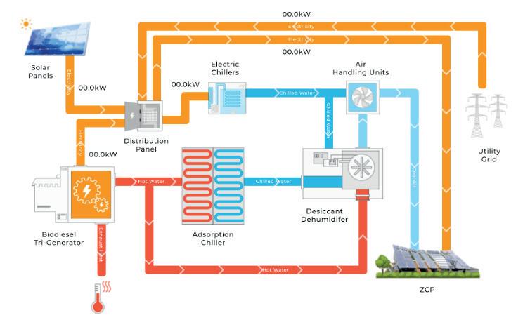
Active Systems
Modular Integrated Construction – MiC is an innovative construction method whereby free-standing integrated modules are manufactured in a prefabrication factory and then transported on site for installation in a building.4
The emMiC system in CIC-ZCP makes use of a stormwater box culvert as the heat extraction and rejection media of the Air-conditioning system.5
In an air-conditioner, hot air in the room is sucked in by a grill located on the bottom of the indoor unit, which then flows through some pipes through which the refrigerants also flow in (the most common refrigerant gases include HFCs R-410A, CFCs such as R-22 and hydrocarbons such as R-290). The refrigerant fluids absorb the heat from the hot air and become hot gases themselves. After passing through the compressor, the hot gas reaches the condenser in the air-conditioner and condenses to cooled liquid. The stormwater, which is relatively low in temperature (about 20 degrees Celsius), serves as an excellent condensing medium for the air conditioner.
71 Chemistry and Biology
Fig. 1 CIC-ZCP energy system
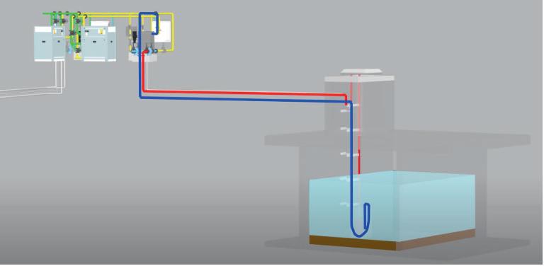
Compared with a typical AC system/electrical heating system, emMiC can reduce energy consumption by 50% and 70% for cooling and heating respectively5 . Using stormwater as a condensing medium also saves fresh water sources, avoids Legionnaire’s disease and mitigates the urban heat island effect*.
*(Urban) Heat island effect: the effect that cause an area of higher temperature relative to surrounding areas, occurs when high concentration of buildings, pavements and other man-made infrastructures absorb and maintain heat radiation.
Air Improvement
Photovoltaic (AIPV) Glass Canopy
CIC-ZCP adopts the Cadmium Telluride nano thin-film photovoltaic technology to generate renewable energy from solar power.
Cadmium Telluride (CdTe) solar cells contain thin-film layers of CdTe material as a semiconductor to convert absorbed solar energy into electricity. This type of solar cell is separated into 5 layers: a copper-doped carbon paste cathode (back content), p-type cadmium telluride and n-type cadmium sulfide (CdS) layers in the middle, a tin oxide or cadmium-based stannous oxide transparent layer acting as the anode (front content), and finally glass substrate on the outside.7
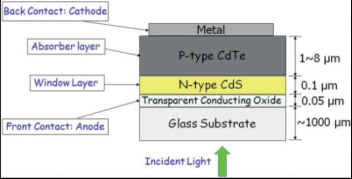 Fig.2 emMiC operation system
Fig.3 Schematic view of CdS/CdTe thin-film solar cell
Fig.2 emMiC operation system
Fig.3 Schematic view of CdS/CdTe thin-film solar cell
72 Scientific Harrovian 2022
8Among all polycrystalline compound semiconductors, CdTe has become a proven TFSC material for its potentiality in several of its advantages and easier methods of thin film depositions:
- An ideal solar cell has a direct band gap of 1.4 eV to absorb the maximum number of photons from the sun’s radiation. CdTe has a near optimum band gap of 1.44 eV, which, compared to the most widely used material – silicon – in the PV industry (indirect band gap of 1.1 eV), is a much more suitable fit in electricity production.8
- CdTe has a high absorption coefficient due to its short absorption length. It absorbs over 90% of accessible photons (hv > 1.44 eV) in a 1 μm thickness.
- The economic cost of producing CdTe cells (associated with polycrystalline and glass) is much lower than the production involving bulk silicon.
- The polycrystalline layers of a CdTe solar cell can be deposited via many different techniques (such as closed-spaced sublimation, physical vapor deposition, RF magnetron sputtering and more).
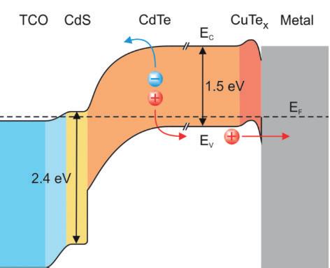
Passive Designs
Cross Ventilated Layout
Cross ventilation is a natural method of cooling the inside of a building —a method that doesn’t require any maintenance cost, carbon emission, or energy consumption, yet still is effective.
The system relies on wind to force external cool air into the building through an inlet (such as a window or wall louver). When this happens, the two sides of the building will be hit with different amounts of pressure. The pressure change will hence force air to the area with lower pressure, which is where the outlet (such as a higher window opening) is located.9 The air circulation allows the inside of the building to experience cool breezes (This is why you will experience breezes when having both the window and room door open).
CIC-ZCP locates its main façade to face the southeast to increase efficiency, as the prevailing summer breeze comes from that direction. Compared to a conventional building, the building’s cross ventilated layout is estimated to have reduced the need for air-conditioning by over 34%.3
Fig. 4 Band energy diagram of the CdS/CdTe solar cell
73 Chemistry and Biology
Wind Catcher
A wind catcher is a small chimney fitted on the roof of the building. As the velocity of wind flowing over the roof is greater than the lower windows of the building, air in the shaft is forced down to cool the building. External air from the roof improves the ventilation potential for areas furthest away from the windows and therefore is not touched on by the cross-ventilation system.
Renewable Energy
Renewable energy is a natural energy that will never deplete and can be used again and again. Many practices were taken to increase the use of renewable energy on site, including but not limited to:
- ZCP has its energy generated mainly from biodiesel. Biodiesel is a type of diesel fuel derived directly from plants and animals, or indirectly from agricultural, industrial, domestic, or commercial waste, and consisting of long chain fatty acid esters. The biodiesel tri-generation system on site is estimated to supply over 129% of ZCP’s energy demand.3
· Different areas of the building are assessed to investigate the irradiance level, and 3 types of pho tovoltaic panels are used based on study. Polycrystalline Silicone PV panels, Building Integrated Photovoltaics, and cylindrical CIGS thin film PV panels are integrated to the inclined main roof, viewing deck, and Air-Tree installation respectively. The PV panels are estimated to produce about 57% of ZCP’s energy demand.3
Contribution to community
Reducing carbon emissions in buildings has a crucial role in achieving the Paris climate goals and achieving net zero emission by 2050. An effective, efficient carbon neutral building takes cost-effective technologies in advantage to reduce emissions. It also serves as an lead example to improve health, equity, and economic prosperity in local communities. The action to invest in carbon neutral buildings around the world remains a priority and symbolises an important step taken in achieving sustainable development.
Bibliography
1 Peng, Changhai. “Calculation of a building’s life cycle carbon emissions based on Ecotect and building information modeling.” ScienceDirect, 20 Jan. 2016, www.sciencedirect.com/science/ article/abs/pii/S0959652615011695.
https://www.sciencedirect.com/science/article/abs/pii/S0959652615011695
2 Sikra, Sonya. “How Does Construction Impact the Environment?” Gocontractor, 21 June 2017,gocontractor.com/blog/how-does-construction-impact-the-environment/.
https://gocontractor.com/blog/how-does-construction-impact-the-environment/
3 CIC-ZCP. “Zero Carbon Park. Our Mission, Vision & Value.” CIC-Zero carbon park main webpage, zcp.cic.hk/eng/mission.
https://zcp.cic.hk/eng/mission
4 “Modular Integrated Construction.” Buildings Department, 28 Oct. 2022, www.bd.gov.hk/en/resources/ codes-and-references/modular-integrated-construction/index.html#:~:text=Modular%20Integrated%20Construction%20(MiC)%20refers,for%20installation%20in%20a%20building.
https://www.bd.gov.hk/en/resources/codes-and-references/modular-integrated-construction/index.html#:~:text=Modular%20Integrated%20Construction%20(MiC)%20refers,for%20installation%20in%20a%20building.
5 Build King Contruction Ltd. “CIC Zero Carbon Park - emMiC (Stormwater Air Conditioning System).” CIC Zero Carbon Park - emMiC (Stormwater Air Conditioning System), e-book ed., p. 1. Pdf.
6 Ashish. “How Does an Air Conditioner (AC) Work?” ScienceABC, 22 Jan. 2022, www.scienceabc.com/ innovation/air-conditioner-ac-work.html.
https://www.scienceabc.com/innovation/air-conditioner-ac-work.html
7 Richariya, Geetam, and Anil Kumar. “Solar cell technologies.” ScienceDirect, 2020, www.sciencedirect.com/topics/engineering/cadmium-telluride-solar-cell.
8 Amin, Nowshad, and Seyed Ahmad Shahahmadi. “Sustainable Energy Technologies & Sustainable Chemical Processes.” ScienceDirect, 2017, www.sciencedirect.com/topics/engineering/ cadmium-telluride-solar-cell.
9 “Cross Ventilation | Wind Effect Ventilation | Moffitt Corp.” Moffitt, www.moffittcorp.com/wind-effect-cross-ventilation/.
Scientific Harrovian 2022
74

Medicinal Applications of
SPIDER SILK
by Jasmine Wong
75 Chemistry and Biology
Silk is a protein fibre spun by spiders to form webs for nests and cocoons, or to catch prey. Their unique properties, such as high toughness and extensibility, make them an excellent new biomaterial.
How is dragline silk naturally produced?
Each orb-weaving female spider has seven different glands, producing seven types of silk with unique properties depending on their purposes. The silk is stored as a liquid in the internal silk glands before it is secreted. It passes through the spigot to the spinnerets on the spider’s abdomen, where it is spun into fibre, forming gossamer [2, 18]. Among these seven different glands, the major ampullate (MA) silk is one of the spider’s most valuable silks, also known as dragline silk.
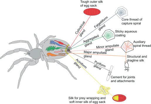
Mechanical Properties of Dragline Silk
The mechanical properties of dragline silk are affected by the amino acids present, insect size, diet, and body temperature [24]; the differences in mechanical properties are dictated by the type of secondary structure of the spider silk, which can be split into four different major motifs [9].
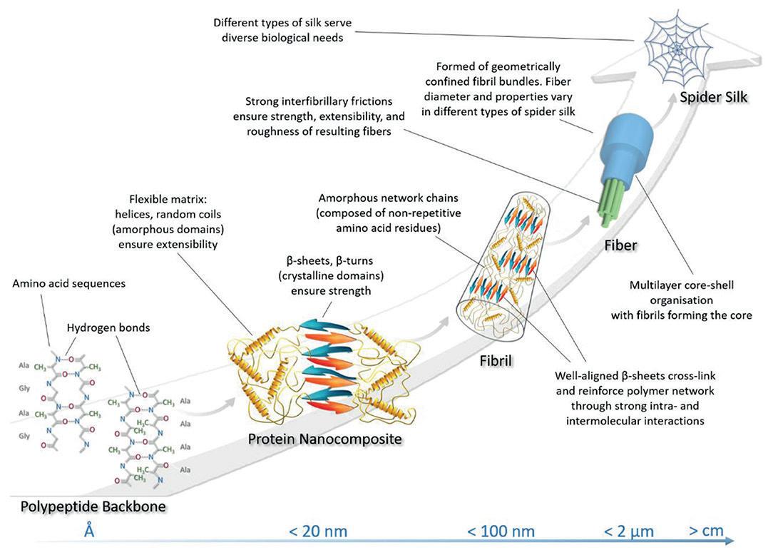 Figure 1: The seven different types of silk produced by Spiders and the different shapes and properties of the silk shown in a diagram [14]
76 Scientific Harrovian 2022
Figure 2: A diagram showing the composition of spider silk with a description of how it aids in causing the properties it possesses [8].
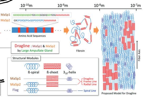
In summary, the primary structure of dragline silk is composed of a sequence of repetitive glycine and alanine blocks [25]. The chain of alanine is mainly found in the crystalline domains of the protein nanofibril composite as shown in Figure 2, whereas glycine is located in the amorphous matrix consisting of beta turns.
High Young’s modulus and tensile strength
The tensile strength (the greatest stress before breaking) of dragline silk is on par with the tensile strength of high-carbon steel (650 Mpa) [19]. The natural dragline silk produced by spiders can have a strength of up to four or five times that of steel. It has one of the highest breaking energies; hence, it is very tough and can withstand a large impact force without breaking [11]. Dragline silk also can undergo large tensile and compressive strains without fracturing or experiencing plastic deformation.
Dragline silk requires a large force of 1.1 x 10⁹ Nm-2 to break under tension when stretched out at either end [9]. To put this into perspective, if you were to be stepped on by an average-sized African bush elephant that weighs 6,000 kg and has a foot size of 0.3 m2 , it would only exert an estimated 20,000 Nm-2 on you. A spider silk would require 55,000 elephants stacked on top of each other to break. If the silk is stretched to its yield point, some of the intermolecular interactions would break, causing the amorphous protein chains to extend, uncoil, and straighten out whilst keeping the polypeptide chain intact. As a result, it can be plastically elongated without losing strength under tensile stress – giving dragline silk its high tensile strength property. Furthermore, the combination of being able to withstand high stress yet experiencing little strain due to untangling of amorphous chains means spider silk has a high Young’s modulus value of 10 GPa. Young’s modulus is the stress-to-strain ratio, i.e., how much pressure will give a certain amount of deformation. This means it is very stiff and resistant to bending or stretching, due to hydrogen bonds breaking by a frictional stick-slip motion, in which energy is dissipated through the amorphous matrix.
Elasticity
The elastic properties of dragline silk further make it an excellent material. The extension of spider silk can be up to two to four times its original length, which is much longer than steel [11]. If an insect flies perpendicular to the silk, the web would need to absorb most of the kinetic energy of the insect’s forward velocity to bring it to a stop without causing the prey to be catapulted out of the web. The dragline silk can do this by transferring 65% of the kinetic energy to thermal energy and storing the rest as elastic deformation as it stretches and recoils back to its original shape and length. To do this, the silk must be extremely tough and extensible. Atomic force microscopy reveals that the silk contains a fibrillar structure with bleated fibrils at the fibre’s core. Bleated fibril is a section of fibre that has a completely non-repetitive random arrangement of loops. This core allows for a large force to be applied without breaking by extending itself,
77 Chemistry and Biology
Figure 3: Molecular breakdown of the protein structure that makes up spider dragline silk [33]
as shown in Figure 4 below. Figure 4 also shows that dragline silk has a toughness of 160 MJm-3, meaning the silk can absorb a large amount of energy per unit volume and recoil after a force has been removed. This is because the surface crack propagates only up to a certain point in the crystallite [9] [24].
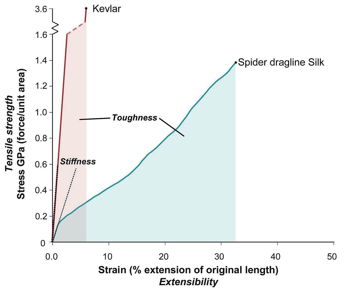
Dragline silk is fibroin and is made up of MaSp1 and MaSp2 Spidrion proteins which are composed of a repeating sequence of approximately 3,500 amino acids with 42% glycine and 25% alanine [6]. Glycine-rich regions give spider silk its elastic properties where a sequence of 5 amino acids is repeated. Unlike other amino acids, which have carbon instead, spider silk contains hydrogen as a side chain [3]. This is further enhanced by the fact that a β turn (180o) occurs periodically after each complete sequence, forming a β-spiral. Furthermore, the β spiral provides a tight helix which can extend with the amorphous area in the silk structure, providing elastic and tough properties. Its toughness is also brought about by the high molecular mass of the protein, causing many Van der Waal forces between the short-chain amino acids as well as the many hydrogen bonds between carbonyl and amide groups.
These forces are embedded in an amorphous glycine matrix consisting of helical and β-turn spiral structures. Additionally, the solid intermolecular forces of attraction between the layers of crystalline structure, i.e. disulfide bridges, give rise to elastic properties. Hence, when a force is applied, the silk is initially slightly stiff until the weak cross-links between the tangled chains are broken. The chains are then uncoiled, increasing strain for a little extra stress until the molecules become aligned. The band now becomes stiff as the strong covalent bonds between the atoms are stretched. On releasing the stress, the chains recoil back to their original structure.
The spring-like structure of the proline provides the corresponding restoring force to balance the torque acting on it [11]. β-turn is also neatly arranged to form a ‘cable-like structure’ which increases the crystalline stability and strengthens the spider silk. The elastic properties are further enhanced by the GPGXX region in the bleated fibrils where the hydrophobic crystalline structure is. This comes from having strong covalent bonds between every three amino acids in the helical structure, which further increase the tensile strength.
The fibre extensibility of the dragline silk compensates for strength when fibres are loaded on long silk, causing the web fibre to bend downwards, stretching the fibre with F=mg/2sinθ. Dragline silk is different from other low extensibility fibres: rather than the deflection angle becoming smaller, which will cause a larger force to develop, a larger angle allows for a higher εmax of 0.27. This is significant compared to the Kevlar fibre because even with a tenth of the normalised deformation, it will only support a load 40% less than the dragline silk. This is effective for spiders when catching insects flying at a high velocity.
78 Scientific Harrovian 2022
Figure 4: A stress-strain graph comparing kevlar and spider dragline silk [29]
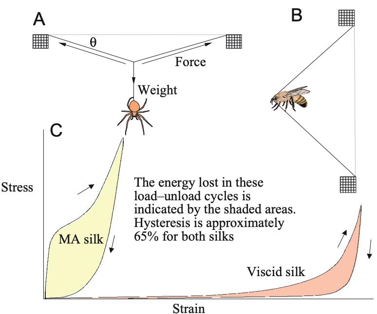
Supercontraction
When submerged in water, spider silk fibres contract, causing a phenomenon known as a super contraction [11]. Changing humidity and moisture can cause the dragline silk to shrink by 40-50% and decrease stiffness by three orders of magnitude, causing it to behave like rubber. Supercontraction, therefore, keeps the web taut. This is believed to be the result of the polar acids forming bonds and interacting with neighbouring atoms.
Biocompatibility
The proteins in dragline silk are biocompatible to humans as they do not contain any toxic substances, nor do they cause immune rejection reactions from the human body. It was suggested in a study that silk produced by the common house spider, T. domestic, had bacteriostatic effects on B. subtilis [32] . The reasons for the reduction were suggested to be due to the copious amounts of glycoproteins in the silk. The hydrophobic nature of both the protein’s crystalline region and the GPGXX sequence can be degraded under specific conditions, and the degraded products can be absorbed by human tissue; hence it is an ideal wound suture and prosthesis-making material.
Glycine acts as an antioxidant, anti-inflammatory, and immunomodulator since it acts as a neurotransmitter in the central nervous system [4]. Being an immunomodulator means that it can change the immune system of the host by activating or suppressing it. Since spider silk is a natural material, it is cheaper and easier to use than manufactured materials. It has antimicrobial properties and is non-immunogenic, non-reactive and biodegradable. The response of mammalian cell lines cultured in vitro to Nephila clavipes further demonstrated that it did not evoke an autoimmune response [32].
Recombinant spider silk production
Harvesting a substantial amount of spider silk from natural sources is a difficult task; hence scientists have been able to produce recombinant spider silk to meet the demands [23]. To form a recombinant protein, recombinant DNA (rDNA) must first be formed. rDNA is a DNA strand that is formed by the combination of two or more DNA sequences from the same species. It can be artificially produced using rDNA technology to put into a host cell. The most common recombinant spider silk proteins are based on the sequence of Araneus diadematus. The most common host organisms used to manufacture the recombinant protein are E.coli, as they proliferate and have high cell density and easy transformation, i.e. high efficiency of introducing DNA molecules into cells and increased ability to express proteins.
Figure 5: Graph of stress-strain to show the properties of Dragline silk, for example, toughness and stiffness
79 Chemistry and Biology
The formation of recombinant protein is as follows:
1. Restriction enzymes cut at a specific sequence of DNA to produce short, single-stranded overhangs[12].
2. Recombinant DNA is formed when the cut DNA fragment (target gene for the production of spider’s silk) is inserted into a vector (a plasmid) using DNA ligase.
3. The resulting vector is inserted into a host cell, ie. E.coli, in a process called transformation. This is done by shocking the bacterial cells with conditions such as high temperature to encourage them to take up foreign DNA.
4. The bacterial cells can now produce the recombinant protein.
5. Once the protein has been produced, the bacterial cells can be split open to release it.
6. The target protein must then be purified or separated from the other contents of the cells by biochemical techniques due to the presence of many other macromolecules around the bacteria besides the target protein.
Medical applications
Tissue engineering
Langer and Vacanti defined tissue engineering as ‘an interdisciplinary field which applies the principles of engineering and life sciences towards the development of biological substitutes that restore, maintain or improve tissue function’ to induce tissue-specific regeneration processes [5]. There are two approaches to tissue engineering. ‘Bottom-up’ is the modular assembly of building units into tissue resembling constructs [15]. ‘Top-down’ is the simple combination of existing components within a given structure.
Example 1: Silk Matrix for tissue-engineered anterior cruciate ligament (ACL)
The high tensile strength and Young’s modulus matches the mechanical properties needed for an ACL [1]. It is also a biocompatible material and avoids bioburdens associated with mammalian-derived materials. It also does not degrade quickly, which means it provides sufficient time for the host tissues to infiltrate and eventually grow back and stabilise the leg.
Example 2: Cardiac regeneration
The primary cause of impaired heart function is the loss of cardiomyocytes [10]. One innovative approach is to use spider silk to help grow new cardiac muscle tissue. By using bioengineered spider silk hydrogels as a base, scientists have been able to restore heart tissues. Cardiomyocytes, harvested from human-induced pluripotent stem cells (hiPSCs), are genetically modified using CRISPR/Cas-9 to allow the surface to be negatively charged, so that it can adhere to the positively charged bioengineered spider silk protein. This allows hypertrophy which repairs the sections of the heart. More importantly, the new cardiomyocytes also demonstrate the ability to communicate with other cells.
Prosthesis
An electro-tendon is a part of the robotic hand made up of spider silk which allows electrical signals to be transmitted to the pressure sensor by the transmission of electrical signals through to the pressure sensor, allowing the finger to bend under the control of a motor. It is able to do this as spider silk is a super tough conductor that can act as an electrode. The durable and flexible nature of spider silk allows the electrotendon to withstand up to 40,000 cycles of bending and stretching in a prosthesis hand [22].
80 Scientific Harrovian 2022
Figure 6: schematics of a prosthetic hand using spider’s silk [22]
Silk optics
Researchers from Taiwan have made biosensors using spider silk [20]. The dragline silk is harvested from Nephilia pilipes which is native and abundant in Taiwan. Spider silk forms the core of the optic fibres, whilst the biocompatible photocurable resin acts as the cladding. It is further enhanced by adding a nanolayer of gold to enhance the fibre’s sensitivity. In optomechanics, spider silk can be used to form sensors that can detect and measure tiny changes in the refractive index of a biological solution which contains glucose [30]. The beam produced is called a photonic nanojet (PNJ). This is useful as it can be used for biomedical nanoimaging applications, such as measuring blood sugar levels with a higher degree of accuracy without being invasive and expensive [20].
Sutures
Biotechnological spider silk production has enabled scientists to genetically modify the sequence of amino acids within the spider silk, thus altering the chemical and physical properties of spider silk proteins. With artificial spider silk, molecules like antibiotics and fluorescent dyes can be attached to desired soluble silk protein [13]. Antiseptic properties effectively clot blood because of its high vitamin K content. Hence, the rapid regeneration of biocompatible and multifunctional spider silk can have a wide range of applications that is useful for biomedical applications.
The point of sutures is to promote wound healing and avoid infections, but the suture itself is susceptible to causing bacterial infection of biofilms [11]. Thus, an extra layer of antibiotic-based antibacterial coating is added to the sutures to prevent bacterial biofilm formation. However, as microorganisms mutate and become resistant to antibiotics due to selection pressure causing evolution by natural selection, an alternative solution to this problem must be found. The biocompatibility, minimal immune response and controlled biodegradability make dragline silk most suitable for this role. Additionally, because spider silk can be easily degraded, it eliminates any changes or dressing removals that would typically be painful for the patient [7].
Conclusion
In conclusion, the dragline silk produced in the major ampullate gland in spiders provides a naturally high Young’s modulus, tensile strength, elasticity and extensibility. It is not only biocompatible and biodegradable, but it also does not evoke an immune response when used on other mammals, making it an attractive material for biomedical applications such as sutures and optic fibres.
However, the source of dragline silk production should be carefully considered, as exploiting spider glands is unethical since it is unnatural for them to keep making silk. In some cases, it is also inhumane to do so as they are involuntarily sedated with carbon dioxide gas and pinned down by their limbs and abdomen to keep them in place. Tweezers are then used to pull out silk from the spinnerets and attach it to the pool with some glue. The motor then begins to spin to harvest the silk from the spider [26].
Alternative methods should be found to avoid harming the spiders. This can be done by studying the structure of the spiders and using biomimicry to replicate the properties.
Figure 7: The basics of a fibre optic cable [28]
81 Chemistry and Biology
Bibliography
[1] Altman, Gregory H., et al. “Silk matrix for tissue engineered anterior cruciate ligaments.” Science Direct, October 2002, https://www.sciencedirect.com/science/ article/abs/pii/S0142961202001564. Accessed 1 November 2022.
[2] Ault, Alicia. “Ask Smithsonian: How Do Spiders Make Their Webs?” Smithsonian Magazine, 3 December 2015, https://www.smithsonianmag.com/smithsonianinstitution/ask-smithsonian-how-do-spiders-make-webs-180957426/. Accessed 28 October 2022.
[3] Betts, M. J., and R. B. Russell. “Glycine: Amino acid properties and consequences of substitutions.” Russell Lab, M R Banes, 2003, http://www.russelllab.org/aas/ Gly.html. Accessed 28 October 2022.
[4] BYJU. “Glycine - Structure, Properties, Uses & Benefits with Images and FAQs.” Byju’s, https://byjus.com/chemistry/glycine-structure/. Accessed 28 October 2022.
[5] Campuzano, Santiago, and Andrew E. Pelling. “Tissue Engineering Approaches in the Design of Healthy and Pathological In Vitro Tissue Models.” Frontiers, 26 June 2017, https://www.frontiersin.org/articles/10.3389/fbioe.2017.00040/full. Accessed 1 November 2022.
[6] CHM BRIS. “Spider Silk.” Spider Silk, http://www.chm.bris.ac.uk/motm/spider/page3.htm. Accessed 28 October 2022.
[7] Cuffari, Benedette. “Role of Spider Silk in Biomedicine.” News Medical, 14 December 2020, https://www.news-medical.net/life-sciences/Role-of-Spider-Silk-inBiomedicine.aspx. Accessed 6 November 2022.
[8] Doistau, Benjamin, et al. “Recent Advances in Development of Functional Spider Silk-Based Hybrid Materials.” Frontiers, 29 May 2020, https://www. frontiersin.org/articles/10.3389/fchem.2020.00554/full. Accessed 12 November 2022.
[9] Ebrahimi, Davoud, et al. “Silk–Its Mysteries, How It Is Made, and How It Is Used.” NCBI, 24 August 2015, https://www.ncbi.nlm.nih.gov/pmc/articles/ PMC4936833/. Accessed 29 October 2022.
[10] Esser, T. U. “Article Highlight: Spider silk could be used to make artificial heart tissues.” Materials Today, 28 June 2021, https://www.materialstoday.com/ biomaterials/news/article-highlight-spider-artificial-heart-tissues/. Accessed 5 November 2022.
[11] Gu, Yunqing, et al. “Mechanical properties and application analysis of spider silk bionic material.” De Gruyter, 24 August 2020, https://www.degruyter.com/ document/doi/10.1515/epoly-2020-0049/html?lang=en#:~:text=The%20main%20chemical%20components%20of,has%20excellent%20elasticity%20and%20 strength.&text=Appearance%20of%20natural%20spider%20silk. Accessed 5 November 2022.
[12] Khan Academy. “Overview: DNA cloning (article).” Khan Academy, 2022, https://www.khanacademy.org/science/ap-biology/gene-expression-and-regulation/ biotechnology/a/overview-dna-cloning. Accessed 1 November 2022.
[13] Kirsh, Danielle. “Artificial spider silk: Why you should care about it.” Medical Design & Outsourcing, 10 January 2017, https://www. medicaldesignandoutsourcing.com/artificial-spider-silk-used-medical-applications/. Accessed 1 November 2022.
[14] Ko, Frank K., and Lynn Y. Wan. Handbook of Properties of Textile and Technical Fibres. 2 ed., The Textile Institute Book Series, 2018.
[15] Langer, Robert. “Tissue Engineering.” Science, 14 May 1993, https://www.science.org/doi/10.1126/science.8493529. Accessed 1 November 2022.
[16] Mahan, Gerald D. “amorphous solid | physics | Britannica.” Encyclopedia Britannica, 31 July 2019, https://www.britannica.com/science/amorphous-solid. Accessed 12 November 2022.
[17] “Mechanical properties and application analysis of spider silk bionic material.” De Gruyter, 2020, https://www.degruyter.com/document/ doi/10.1515/epoly-2020-0049/html?lang=en#:~:text=The%20main%20chemical%20components%20of,has%20excellent%20elasticity%20and%20 strength.&text=Appearance%20of%20natural%20spider%20silk. Accessed 28 October 2022.
[18] Merda, Chad. “Nature curiosity: How do spiders make silk?” Forest Preserve District of Will County, 4 October 2019, https://www.reconnectwithnature.org/ news-events/the-buzz/nature-curiosity-how-do-spiders-make-silk. Accessed 28 October 2022.
[19] Nuclear Power. “High-carbon Steel | nuclear-power.com.” Nuclear Power, 2022, https://www.nuclear-power.com/nuclear-engineering/metals-what-are-metals/ steels-properties-of-steels/high-carbon-steel/. Accessed 28 October 2022.
[20] Optica. “Researchers create biosensor by turning spider silk into optical fiber | News Releases.” Optica, 2 August 2022, https://www.optica.org/en-us/about/ newsroom/news_releases/2022/august/researchers_create_biosensor_by_turning_spider_sil/. Accessed 5 November 2022.
[21] Orkin. “What is Spider Silk? | Spider Silk Types & Facts.” Orkin, 2022, https://www.orkin.com/pests/spiders/spider-silk-facts-and-information. Accessed 28 October 2022.
[22] Pan, Liang, et al. “A supertough electro-tendon based on spider silk composites.” Nature communications, 12 March 2020, https://www.nature.com/articles/ s41467-020-14988-5#:~:text=The%20electro%2Dtendon%20is%20a,without%20any%20change%20in%20conductivity. Accessed 1 November 2022.
[23] Salehi, Sahar, et al. “Spider Silk for Tissue Engineering Applications - PMC.” NCBI, 8 February 2020, https://www.ncbi.nlm.nih.gov/pmc/articles/ PMC7037138/. Accessed 1 November 2022.
[24] Saravanan, D. “Spider Silk - Structure, Properties and Spinning.” Journal Of Textile and Apparel, Technology and Management, vol. 5, no. 1, 2006, p. 1. JTATM. Accessed 29 October 2022.
[25] Shishir, Hasan. “Spider silk.” SlideShare, 13 May 2014, https://www.slideshare.net/sheshir/spider-silk-34606555. Accessed 6 November 2022.
[26] “Spider Silk Harvesting.” jwz.org, 2014, https://www.jwz.org/blog/2015/03/spider-silk-harvesting/. Accessed 6 November 2022.
[27] “Tissue Engineering.” Science, 14 May 1993, https://www.science.org/doi/10.1126/science.8493529.
[28] Vesselnet. “Three Basic Components of a Fiber Optic Cable.” Vesselnet, https://www.vesselnetintegrated.com/single-post/three-basic-components-of-a-fiber-opticcable. Accessed 12 November 2022.
[29] Vincentsarego. “File:Wikipedia Kevlar Silk Comparison.jpg.” Wikimedia Commons, 17 June 2011, https://commons.wikimedia.org/wiki/File:Wikipedia_ Kevlar_Silk_Comparison.jpg. Accessed 12 November 2022.
[30] Weiler, Sarah. “Spider silk spins a stronger lens | Post Scripts | Sep/Oct 2020 | BioPhotonics.” Photonics.com, October 2020, https://www.photonics.com/Articles/ Spider_silk_spins_a_stronger_lens/a66044. Accessed 5 November 2022.
[31] Whittall, Dominic R., et al. “Host systems for the production of recombinant spider silk.” Cell, 10 October 2020, https://www.cell.com/trends/biotechnology/ fulltext/S0167-7799(20)30244-4. Accessed 1 November 2022.
[32] Wright, Simon. “Evidence for antimicrobial activity associated with common house spider silk.” NCBI, 25 June 2012, https://www.ncbi.nlm.nih.gov/pmc/ articles/PMC3443048/. Accessed 1 November 2022.
[33] Zhao, Yue. “File:0.Figure.png.” Wikimedia Commons, 7 June 2016, https://commons.wikimedia.org/wiki/File:0.Figure.png. Accessed 12 November 2022.
[34] CRISPR. “CRISPR/Cas9 | CRISPR.” CRISPR Therapeutics, 2022, https://crisprtx.com/gene-editing/crispr-cas9. Accessed 14 December 2022.
82 Scientific Harrovian 2022

The Power of STEM CELLS
by Jais Brar
83 Chemistry and Biology
As more research has been conducted in pursuit of new medical advances, stem cell therapy has emerged as a promising and hopeful treatment method for many conditions. In 2021 alone, the stem cell market was worth 77 billion HKD, and is continually growing at an exponential rate, it is estimated to surpass 246 billion HKD by 2030. (1) Stem cells can be used in numerous ways as they have the potential to develop into any cell found in the body. Unlike most cells that are only able to undergo a limited number of divisions before their death, stem cells can undergo an infinite number of divisions (2), making them one of the most unique scientific discoveries in the past 100 years.
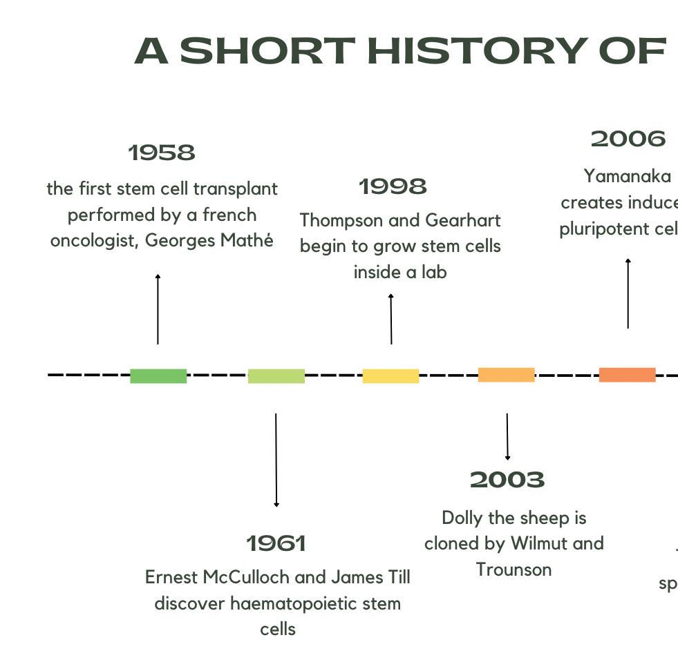
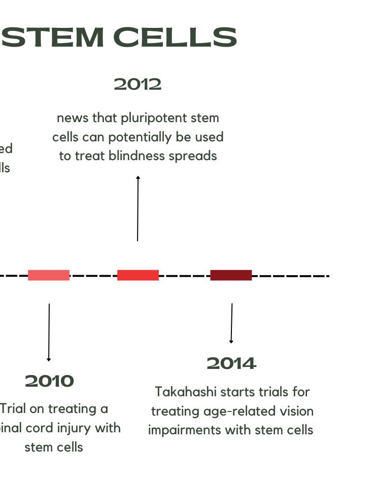
The History of Stem Cells
Around 30 years ago, scientists discovered ways to obtain embryonic stem cells from early stage mice embryos. Biologists that studied these cells soon realized that the cells were pluripotent, meaning they have the potential to be converted into any adult body cell. Further laboratory experiments enabled scientists the ability to extract embryonic cells from humans and grow them in lab conditions. Prior to this, in the 1950s, the first stem cell transplant was given to nuclear researchers who had been exposed to radiation from cells found in the bone marrow. This procedure was carried out by Georges Mathé, a French oncologist.
Then later, in the 1960s, Ernest McCulloch and James Till gained new insight into blood cell formation through the discovery of haematopoietic stem cells. (3) This was done by injecting bone marrow cells into mice that had endured nuclear radiation. The cells were seen to develop into the three primary components of blood: red blood cells, white blood cells and platelets. Although these occurrences were rare,it demonstrated that differentiation was possible in bone marrow cells.(4)
Another major breakthrough in the stem cell world was the cloning of the infamous Dolly the sheep. Ian Wilmut and Alan Trounson, at Roslin Institute, managed to clone her using the DNA from an adult sheep’s mammary gland (found in the breast tissue), showing that if an adult cell was fused with an empty egg cell it would mirror the genetic makeup of the body it is being inserted into and replicate all tissues and organs, along with proving that the entire DNA of an adult was present in a single cell. (5) In around 1998, James Thomson and John Gearhart began to grow stem cells in the lab and in 2006, Shinya Yamanaka created induced pluripotent stem cells by injecting four key genes. Stem cells were increasingly used in medical therapy in the early 2000s, such as the first authorized trial on treating someone with a spinal cord injury with stem cells derived from embryos.
84 Scientific Harrovian 2022
This was followed by promising news, in 2012, stating that pluripotent stem cells could potentially be used to treat blindness. Human trials for this began in 2014, with Masayo Takahashi leading the study in treating age-related blindness and easing symptoms.(6) The inserted stem cells differentiated into retinal pigment epithelial cells, which are e damaged or lost in people with vision impairments, therefore improving vision and sight. Another way in which stem cells can be used in treating vision impairments is by collecting corneal limbal stem cells from a donor and using these to treat corneal damage. Although there are not cures for all of the sight related diseases, new remedies relating to optical issues such as glaucoma and wet macular degeneration, are being researched into extensively. (7)
Stem Cell Potencies
Stem cells all have the possibility of turning into specialized cells, however, within stem cells there are different levels of capabilities of transforming into different cell lineages. These include; totipotent, pluripotent, multipotent, oligopotent and unipotent, as shown in Figure 1.
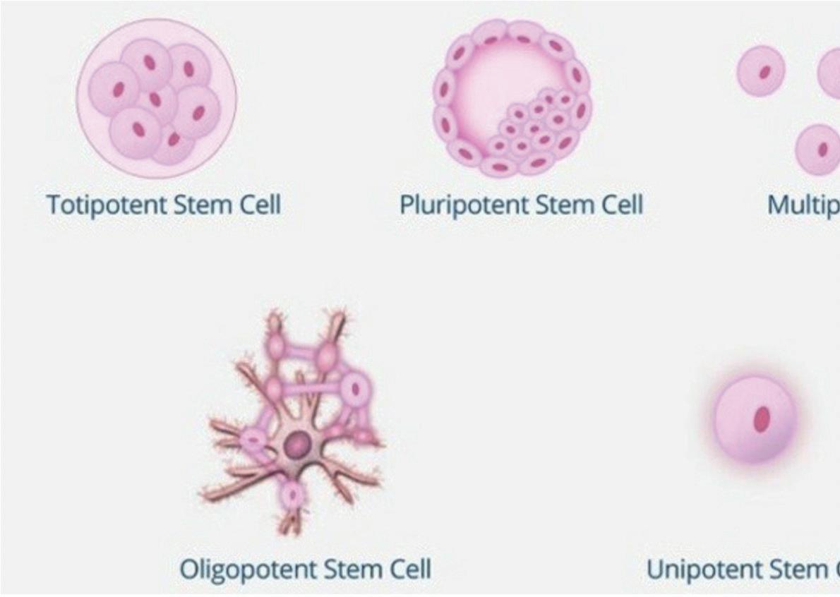

Totipotent Cells
Totipotency is the highest level of potency a stem cell can have. This means that they are able to differentiate into any cell, whether it be embryonic or adult. They can develop into over 200 different types of specialized cell in the body, making their differentiation potential optimal for medical use. Totipotent cells can be extracted from human zygotes, or zoospores, asexually reproductive structures found in algae and fungal species. The zygote is totipotent as it can evolve into any of the three germ layers; ectoderm (which forms the exoskeleton), the mesoderm (which forms the organs), and the endoderm (which forms the inner lining of organs).(9) Despite the fact that these cells fall into the most favorable category of stem cells, they are rarely used in scientific research as not only they are arduous to obtain but also go against various ethical guidelines. (10) As totipotent cells are derived from the first stage of formation of a fetus, obtaining them harms the blastocyst on the sixth or eighth day of development. This in turn destroys the embryo. However, as the embryos used in this practice are donated from IVF clinics and are deemed unwanted, it can be argued that it does not in fact breach any ethical regulations.
85 Chemistry and Biology
Figure 1 – shows the different potencies within stem cells (8)
Pluripotent Cells
After approximately 4 days, the zygote grows into an embryo. Embryonic stem cells are stem cells obtained from the inner cell mass of a blastocyst, which is a human embryo between 3 to 5 days old and contains about 150 cells only. Pluripotent cells can progress into any of the three germ layers, as shown in Figure 2, but not into any extra-embryonic structures, such as the placenta. These types of stem cells are completely pluripotent as they have the potential to be specialized into any body cell and even undergo mitosis, enabling them to divide into larger quantities of stem cells. This trait, along with being easier to acquire, allows them to be greatly useful for medical practices, especially in regenerating organs, tissues and aiding in overcoming a disease with limited treatment availabilities. (10)
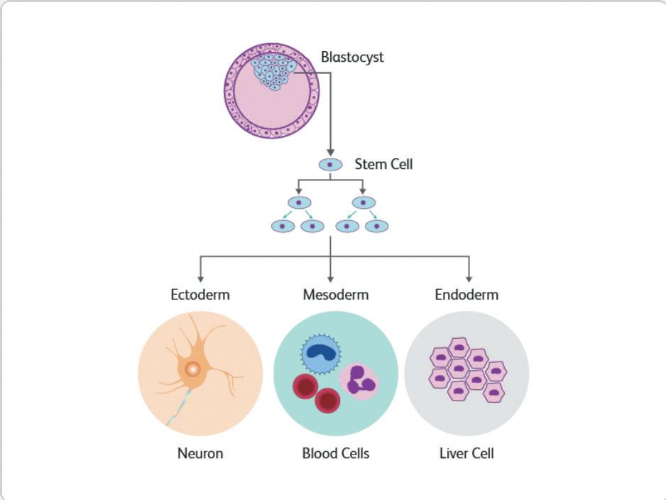
Multipotent and oligopotent cells
These types of cells have a lower differentiation potential compared to pluripotent cells. Multipotent cells are limited in their capacities, they can specialize into sub-cells in a specific cell lineage but cannot develop into any type of cell. An example of multipotent cells are hematopoietic cells, which can be found in bone marrow or in the blood from the umbilical cord. Hematopoietic cells have the ability to evolve into any blood cell but are unable to produce cells that are not blood cells. In recent years, scientists have located the presence of multipotent hematopoietic cells in the heart, which have the tendencies to develop into heart muscle of endothelial cells. These can be extremely helpful in treating blood cancers like leukemia. (12) Oligopotent cells, found within specific tissue e.g. the cornea , can self-renew and regenerate into more lineages within a particular type of cell. They can also form terminally differentiated cells of a specific tissue, meaning oligopotent cells no longer have the ability to undergo mitosis and proliferate. (13)
86 Scientific Harrovian 2022
Figure 2 –This shows the three germ layers that an embryonic stem cell or other pluripotent stem cells can specialize into. (11)
Unipotent stem cells are characterized by their narrow capabilities in cell differentiation. They are only adequate for one singular cell lineage, for example muscle cells only being able to evolve into mature muscle cells. (13) Unipotent cells are distinguished from non-stem cells by a few key characteristics, one of which is being able to self-renew. (14) This is the process in which stem cells can divide and reproduce asexually with the help of mitosis, the cells self-divide in their undifferentiated states, which allow them to be highly beneficial. (15)
Induced pluripotent stem cells
Stem cells can also be induced, meaning they can be created in lab conditions. Induced pluripotent stem cells (iPS) can be derived from adult body cells and have been reprogrammed through inducing genes to make them pluripotent. Like many other scientific advances, these cells were first found in mice fibroblasts (a lineage of cells that are responsible for the production of connective tissue) in 2006 by Yamanaka and then later in 2007 they were first independently produced from human fibroblasts. iPS cells behave in similar ways to embryonic stem cells and hold a lot of the same characteristics, some of which include expression of Embryonic Stem cell markers, chromatin methylation patterns (methylation inhibits gene expression in cells by affecting chromatin structure. Chromatin is a mixture of DNA and special proteins, such as histones) (16), embryoid body formation (three-dimensional aggregates) (17) , pluripotency and many more.
Somatic cells can be reprogrammed into iPS cells by the transcription genes Oct4 (octamer-binding transcription factor 4, a molecular marker for germ cell tumors) (18), Sox2 (SRY-box 2, a marker for multipotential neural stem cells) (19), Klf4 (kruppel-like transcription factor, a protein coding gene) (20) and c-Myc (MYC proto-oncogene, BHLH transcription factor, a regulator of cellular metabolism and proliferation) (21). These factors, along with a few others,can reprogram adult somatic cells (any body cell that is not a gamete) and transdifferentiate them into neural stem cell like structures (22). iPS cells can be an excellent alternative to embryonic stem cells as they are comparatively less invasive, making them more ethical. They can also be especially useful in regenerative medicine (involved with regenerating tissues or organs), disease modeling and drug discovery. (23)
Proliferation and specialization
As mentioned previously, the most prominent attribute of stem cells is that they are able to differentiate into any desired cell. When a cell divides by mitosis, it forms two identical daughter cells and these cells can either: both remain as stem cells, both differentiate into new cells or have one remaining as a stem cell and the other differentiating . When both daughter cells either differentiate or remain as stem cells, it is called a symmetric division, whereas when they both carry out different processes this is known as asymmetric division. The chances of a symmetric division happening, 70%, is relatively high compared to those of an asymmetric division, 30%. This property allows the cells to retain their ability to perpetually divide as it ensures that stem cell levels are kept at a constant level, i.e. stem cells don’t deplete.
When it comes to the mechanics behind the specialization of stem cells scientists are still unsure as to what aids it since they are still relatively new. Producing mature neurons is pivotal to understanding the physiology of human neurons and glial cells and the pathology of neurological diseases, such as epilepsy. The cells differentiate into neural progenitor cells by embryoid body formation, then they are directed into specific neurons. These steps are all accomplished with the help of transcription factors.
Pancreatic β-cell helps in glucoregulation, when blood glucose levels are too high they produce insulin therefore the production and transplantation of these β-cells can treat diabetic patients. For this cells are induced into a definitive endoderm (one of the three germ layers) and then they further differentiate into pancreatic cells. (24) Pluripotent stem (iPS) cells can also be used in myogenic regeneration of skeletal muscle and cardiac muscle, along with hepatocyte differentiation (hepatocytes are parenchymal cells in the liver that are crucial in metabolic action, detoxification and protein synthesis) (25).
Unipotent
87 Chemistry and Biology
Medicinal application of stem cells
Since stem cells are so versatile, they can be used profusely in treating medical conditions. Some of the conditions include:
Tissue regeneration:
Prior to the discovery of pluripotent stem cells, patients with damaged organs or tissues had to wait extensive periods to receive a transplant but in present times scientists can use cell differentiation to grow organs and tissues to replace the organ.(26)The human skin has remarkable regenerative capabilities, especially with the epidermis (the outermost layer of skin) being able to continually regenerate. This is most helpful when treating severe burns and other chronic wounds. Plastic surgeons use stem cells extracted from beneath the skin surface to grow new skin tissue. This newly grown skin tissue is placed onto the open wound, allowing the skin to grow back, closing any exposed wounds. (27)
Cardiovascular disease treatment:
With ischemic heart disease being the leading cause of death in the world, it is essential that there are successful therapies to prevent myocardial infarctions (heart attacks). Scientists have found methods to insert stem cells into the cardiac tissue with a catheter to help regenerate the deteriorated tissue, improving the patient’s quality of life. (28)
Brain disease treatment:
Stem cells can be used to replace and regenerate impaired brain cells that cause neurological disorders, such as Parkinson’s and Alzheimer’s. Parkinson’s disease, for example, is caused by lowered levels of dopamine in the brain due to a loss of nerve cells in the substantia nigra (located within the midbrain). (29) Stem cells can be directly placed in the brain where the damaged nerve cells are, where they regenerate into nerve cells and regulate dopamine levels, suppressing certain symptoms of the disease. Although this is not a definite cure for the disease it can help lessen the symptoms, and hopefully with more development can act as a permanent cure. (30)
Blood disease treatments:
Blood diseases, such as leukemia, multiple myeloma and lymphoma, can be a consequence of genetics, side effects of medication or due to a lack of particular nutrients. (31) The bone marrow produces all blood cells, thus by taking hematopoietic stem cells from the bone marrow or the umbilical cord can produce healthy red blood cells to carry oxygen and healthy white blood cells to fight the cancerous cells, infections and other pathogens. (32) In cases of hemophilia A, a blood disease caused by the lack of blood clotting factor VIII that causes excessive bleeding as the blood isn’t able to clot. Stem cells can differentiate into platelets to aid in blood clotting as a treatment for the disease. (33)
Conclusion
In conclusion, stem cells are exceedingly beneficial to the medical world due to their versatility. Although their use is controversial as they are derived from living organisms and can be seen as wasteful, the amount of effort and money that goes into the research and advancement of stem cells will in turn change the way we perceive them, and reveal how valuable stem cells are to humans.
88 Scientific Harrovian 2022
1. stem-cell-therapy-market-size-share-growth-forecast-2022-2030 [Internet]. [cited 2022 Nov 7]. Available from: https://www.biospace.com/ article/stem-cell-therapy-market-size-share-growth-forecast-2022-2030/
2. Stem-Cell-Research-Quick-Guide.pdf [Internet]. [cited 2022 Nov 9]. Available from: https://research.ucdavis.edu/wp-content/uploads/StemCell-Research-Quick-Guide.pdf
3. the-beginnings-of-stem-cell-therapy [Internet]. [cited 2022 Nov 7]. Available from: https://the-dna-universe.com/2021/06/24/thebeginnings-of-stemw-cell-therapy/#:~:text=Stem%20cell%20therapy%20%E2%80%93%20The%20beginning&text=In%20the%20early%20 1960s%2C%20Ernest,series%20of%20experiments%20in%20mice
4. PIIS002561962100063X.pdf.
5. 20-years-after-dolly-the-sheep-led-the-way-where-is-cloning-now [Internet]. [cited 2022 Nov 7]. Available from: https://www. scientificamerican.com/article/20-years-after-dolly-the-sheep-led-the-way-where-is-cloning-now/
6. dn24970-stem-cell-timeline-the-history-of-a-medical-sensation [Internet]. [cited 2022 Nov 7]. Available from: https://www.newscientist.com/ article/dn24970-stem-cell-timeline-the-history-of-a-medical-sensation/
7. stem-cell-therapy-for-vision-loss [Internet]. [cited 2022 Nov 7]. Available from: https://www.fightingblindness.ca/resources/stem-cell-therapyfor-vision-loss/#:~:text=Stem%20cells%20(at%20the%20top,because%20of%20blinding%20eye%20disease.
8. types-of-stem-cells [Internet]. [cited 2022 Nov 9]. Available from: https://bioinformant.com/types-of-stem-cells/
9. multiomics-mammalian-primary-germ-layers [Internet]. [cited 2022 Nov 7]. Available from: https://www.ebi.ac.uk/about/news/researchhighlights/multiomics-mammalian-primary-germ-layers/#:~:text=endoderm%2C%20and%20ectoderm.-,Three%20primary%20germ%20 layers,%3A%20ectoderm%2C%20mesoderm%20and%20endoderm.
10. s13287-019-1165-5 [Internet]. [cited 2022 Nov 7]. Available from: https://link.springer.com/article/10.1186/s13287-019-1165-5
11. stem-cells [Internet]. [cited 2022 Nov 9]. Available from: https://www.bdbiosciences.com/en-us/learn/research/stem-cells#Overview
12. difference-between-pluripotent-and-multipotent-stem-cell [Internet]. [cited 2022 Nov 7]. Available from: http://www.differencebetween.net/ science/health/difference-between-pluripotent-and-multipotent-stem-cell/
13. 345615 [Internet]. [cited 2022 Nov 7]. Available from: https://www.karger.com/article/pdf/345615#:~:text=Oligopotent%20Cells%20 Oligopotent%20stem%20cells,and%20conjunctival%20cells%20%5B41%5D%20.
14. 10a.%20Stem%20Cells%20-%20Teacher%20Background.pdf [Internet]. [cited 2022 Nov 7]. Available from: https://www.ohsu.edu/sites/ default/files/2019-02/10a.%20Stem%20Cells%20-%20Teacher%20Background.pdf
15. 19575646 [Internet]. [cited 2022 Nov 7]. Available from: https://pubmed.ncbi.nlm.nih.gov/19575646/#:~:text=Self%2Drenewal%20is%20 the%20process,depending%20on%20the%20stem%20cell.
16. Hashimshony T, Zhang J, Keshet I, Bustin M, Cedar H. The role of DNA methylation in setting up chromatin structure during development. Nat Genet. 2003 Jun;34(2):187–92.
17. NBK424234 [Internet]. [cited 2022 Nov 9]. Available from: https://www.ncbi.nlm.nih.gov/books/NBK424234/#:~:text=Embryoid%20 bodies%20(EB)%20are%20the,specific%20cell%20lineages%20from%20PSCs.
18. PubMed entry [Internet]. [cited 2022 Nov 9]. Available from: http://www.ncbi.nlm.nih.gov/pubmed/17205510
19. Ellis P, Fagan BM, Magness ST, Hutton S, Taranova O, Hayashi S, et al. SOX2, a persistent marker for multipotential neural stem cells derived from embryonic stem cells, the embryo or the adult. Dev Neurosci. 2004 Aug;26(2–4):148–65.
20. carddisp.pl [Internet]. [cited 2022 Nov 9]. Available from: https://www.genecards.org/cgi-bin/carddisp.pl?gene=KLF4
21. carddisp.pl [Internet]. [cited 2022 Nov 9]. Available from: http://genecards.org/cgi-bin/carddisp.pl?gene=MYC
22. cr2011107 [Internet]. [cited 2022 Nov 9]. Available from: https://www.nature.com/articles/cr2011107#citeas
23. Singh VK, Kalsan M, Kumar N, Saini A, Chandra R. Induced pluripotent stem cells: applications in regenerative medicine, disease modeling, and drug discovery. Front Cell Dev Biol. 2015;3:2.
24. Oh Y, Jang J. Directed Differentiation of Pluripotent Stem Cells by Transcription Factors. Mol Cells. 2019 Mar 31;42(3):200–9.
25. cmi201597 [Internet]. [cited 2022 Nov 9]. Available from: https://www.nature.com/articles/cmi201597#:~:text=Hepatocytes%2C%20 the%20major%20parenchymal%20cells,by%20secreting%20innate%20immunity%20proteins.
26. 323343 [Internet]. [cited 2022 Nov 14]. Available from: https://www.medicalnewstoday.com/articles/323343#donating-and-harvesting
27. S0022202X15357687 [Internet]. [cited 2022 Nov 10]. Available from: https://www.sciencedirect.com/science/article/pii/ S0022202X15357687
28. repairing-the-heart-with-stem-cells [Internet]. [cited 2022 Nov 14]. Available from: https://www.health.harvard.edu/heart-health/repairingthe-heart-with-stem-cells#:~:text=Stem%20cell%20therapy%20for%20the,help%20regenerate%20damaged%20heart%20tissue.
29. causes [Internet]. [cited 2022 Nov 14]. Available from: https://www.nhs.uk/conditions/parkinsons-disease/causes/#:~:text=Parkinson’s%20 disease%20is%20caused%20by,producing%20a%20chemical%20called%20dopamine.
30. stem-cell-therapy-for-parkinsons [Internet]. [cited 2022 Nov 14]. Available from: https://www.healthline.com/health/parkinsons/stem-celltherapy-for-parkinsons#potential-benefits
31. blood-diseases [Internet]. [cited 2022 Nov 14]. Available from: https://www.niddk.nih.gov/health-information/blooddiseases#:~:text=Many%20blood%20diseases%20and%20disorders,bleeding%20disorders%20such%20as%20hemophilia.
32. blood-disorders [Internet]. [cited 2022 Nov 14]. Available from: https://www.childrenshospital.org/research/programs/stem-cell-programresearch/conditions/blood-disorders
33. hemophilia-treatment-stem-cells [Internet]. [cited 2022 Nov 14]. Available from: https://bioinformant.com/hemophilia-treatment-stemcells/#:~:text=Embryonic%20stem%20cells%20may%20have,embryonic%20stem%20cells%20(ESCs).
89 Chemistry and Biology
Bibiliography

the mechanism of
PHOTOSYNTHESIS
by Gloria Siu
90 Scientific Harrovian 2022
What is photosynthesis?
During our time at school, we have been taught that photosynthesis converts light energy into chemical energy, as demonstrated in the chemical equation: 6CO2 + 6H2O —> C6H12O6 + 6O2. However, the process of how light energy is transformed into chemical energy is more complex than what this simple equation suggests. This article aims to explore the intricacies of photosynthesis and the processes involved in it, revealing that there is more to this fundamental process.
The function of chloroplast and chlorophyll
Chloroplasts are organelles present within plant cells; they serve as the primary site of photosynthesis. Chloroplasts contain a green pigment called chlorophyll, which gives it its green hue. The primary function of chloroplasts is to absorb light energy and convert it into chemical energy [1]. There are two common types of chlorophyll, chlorophyll a and chlorophyll b. While all photosynthetic organisms require chlorophyll a, chlorophyll b is only found in green algae and certain plants [2]. Chlorophyll a absorbs violet-blue and orange-red light from the light spectrum, with absorption occurring within the range of 430 nm to 660nm. In contrast, chlorophyll b only absorbs orange-red light from the light spectrum, with absorption taking place between 450 nm to 650nm [3]. The light energy absorbed excites the electrons in the chlorophyll which is located in the photosystem, this causes it to move through the electron transport chain. - This is shown in Figure 1.
Structure of chloroplast
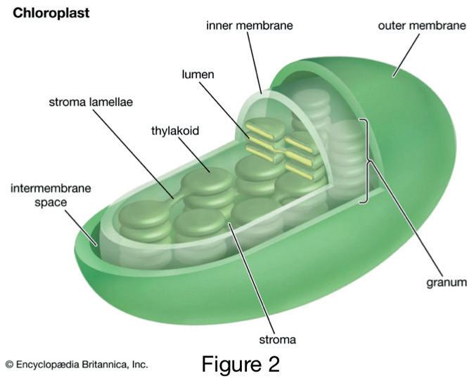
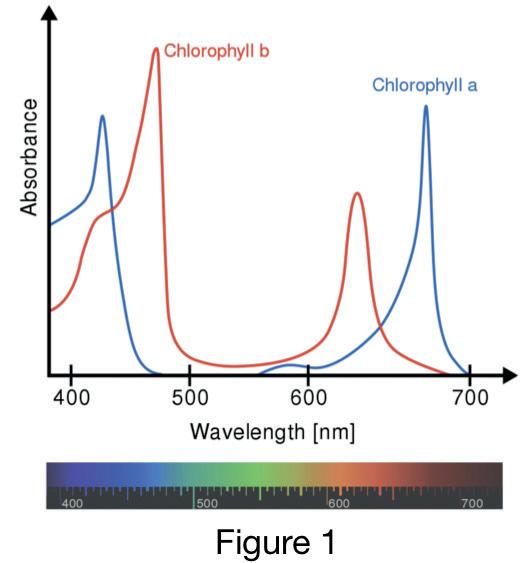
Chloroplasts are made up of many different structures which are shown in Figure 2. The chloroplast is enclosed by a double membrane, and the space between the two membranes is referred to as the chloroplast envelope or intermembrane space [4]. The double membrane surrounding the chloroplast regulates the movement of substances in and out of the chloroplast, thereby controlling the entry and exit of molecules. The granum is a stack made up of disclike structures called thylakoids that are connected by the stroma lamellae. Thylakoids contain molecules of chlorophyll and serve as the site of light-dependent reactions (LDR), the initial phase of photosynthesis. During this stage, light energy is transformed into chemical energy in the form of Adenosine Triphosphate (ATP) and Nicotinamide adenine dinucleotide phosphate (NADPH). Both ATP and NADPH function as energy storage molecules, and they are crucial for the second phase of photosynthesis, known as the light-independent reactions (LIR), which are part of the Calvin cycle [6].
Photosynthetic pigments
Another important photosynthetic pigment other than chlorophyll a and b, which makes up the chloroplast, are carotenoids. Carotenoids absorb light in the blue - green light region in the light
91 Chemistry and Biology
spectrum [28]. They are one of the pigments that gives fruits its colouration, an example of a carotenoid would be β-Carotene (Beta carotene). It can be found in oranges and carrots, giving them their orange colour [7]. Carotenoids also act as antioxidants, as they can protect cells from damage from free radicals [24]. - Figure 3 shows the structure of β-Carotene.
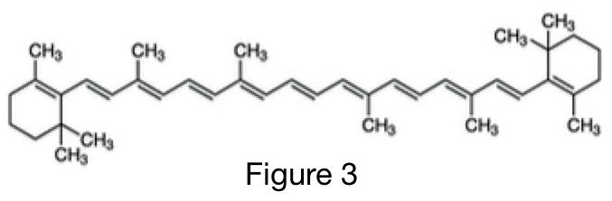
Carotenoids are photosynthetic pigments found in chloroplasts, they are often attached to proteins or membranes [8]. They absorb light in the violet, blue and green regions of the visible light spectrum with wavelengths ranging from around 450 nm to 500 nm [9]. Carotenoids reflect yellow, orange and red wavelengths, giving plants and fruits their yellow or orange-red colours [9]. - The absorption of β-Carotene, compared to chlorophyll a and chlorophyll b, is shown in Figure 4.
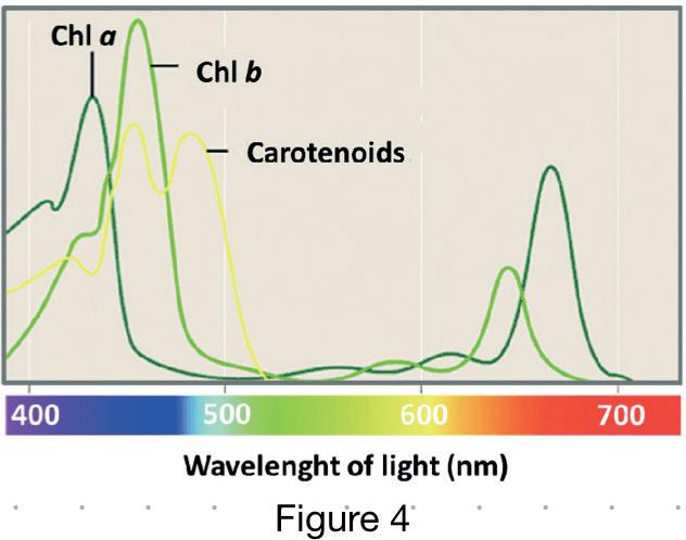
Reactions in photosynthesis
Light-dependent reactions (LDR)
Light-dependent reactions, which are responsible for converting light into chemical energy, take place in the thylakoid membranes located in chloroplasts [6]. There are two photosystems which aid the conversion of light energy into chemical energy; they are known as photosystem I (PSI) and photosystem II (PSII). Photosystems are made up of large complexes of proteins and photosynthetic pigments which aid the absorption of light in the two chlorophyll complexes (Chlorophyll a and chlorophyll b) [10]. The two photosystems are embedded within the thylakoid membrane. - Figure 5 shows this.
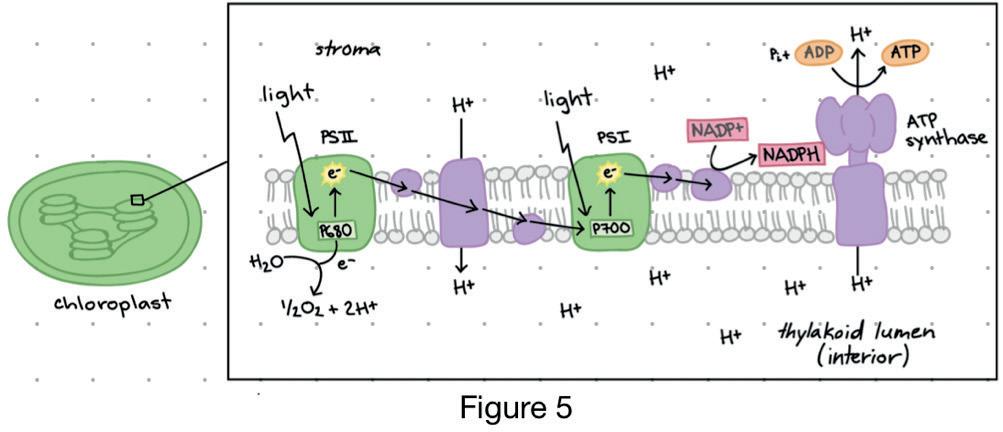
92 Scientific Harrovian 2022
Photosystem II (PSII)
Photosystem II (PSII) is the first photosystem to be reached by light, it contains chlorophyll b, it absorbs light at around 680nm. Its reaction centre is known as P680 due to its optimal absorption of light at this wavelength. Only non-cyclic phosphorylation occurs in PSII [11].
When a photon of light reaches PSII, the energy is transferred to P680, causing the electrons in chlorophyll b to become excited and move to a higher energy level [12]. These electrons travel through the electron transport chain to PSI (A lower energy state compared to PSII), losing energy in the process and leaving a P680+ ion.
The electron carriers found in the thylakoid membrane transport the electrons through the electron transport chain [29]. Photolysis is the breakdown of water using light energy; electrons lost in PSII (after excitation) are replenished with electrons produced from water after photolysis. Oxygen and hydrogen ions (H+) are by-products of photolysis. The movement of electrons in the transport chain releases energy, this energy is used to pump the hydrogen ions (H+) into the thylakoid space. H+ ions contribute to the production of ATP through activation of ATP synthase. When H+ ions are accumulated in the thylakoid space, it diffuses through ATP synthase and energy is released. This energy is used to synthesise ATP through adding ADP to Pi (inorganic phosphate group), this is known as phosphorylation. The ATP synthesised is later used in the light-independent reactions (LIR), as part of the Calvin cycle [10, 17].
Photosystem I
Photosystem I (PSI) involves chlorophyll a, which absorbs light at a wavelength of approximately 700nm, with its reaction centre referred to as P700 [13]. When PSI is excited by a photon of light, an electron in P700 is elevated to a higher energy state and passes through the electron transport chain [13]. The electron is then transferred to a protein known as Ferredoxin (Fd), which is located in the stroma of the thylakoid membrane [14, 15]. Ferredoxin NADP+ oxidoreductase (FNR) is an enzyme that catalyses the transfer of electrons from Ferredoxin to NADP+. The H+ ions that are pumped from the thylakoid space to the stroma as well as the transferred electrons from PSII reduces NADP+ to NADPH [16].
The ATP and NADPH generated from both photosystems are involved in the production of glucose in the Calvin cycle [17]. When ATP is hydrolysed, energy is released by converting ATP to ADP (Adenosine diphosphate) [18]. A phosphate atom is released, providing energy during the LIR (Light independent reactions) [17]. NADPH is converted to NADP+ during the Calvin cycle, and a hydrogen atom is released [17].
Photophosphorylation
There are two types of photophosphorylation, they are known as cyclic and non-cyclic photophosphorylation, as described earlier in the explanation of the two photosystems [25]. Cyclic photophosphorylation only involves PSI. When light reaches PSI, it excites the electrons in P700, the reaction centre of PSI, causing them to move to a higher energy level [13]. The excited electrons will move through the electron transport chain, providing energy [17] which enables H+ ions to be pumped into the thylakoid space. This leads to the activation of ATP synthase which results in the synthesis of ATP. Subsequently, the excited electrons will return to PSI, and this cycle continues to produce more ATP [14].
Both PSI and PSII are involved in non-cyclic photophosphorylation [25]. Excitation of electrons in the P680 reaction centre occurs when light energy is present, which initiates non–cyclic photophos-
93 Chemistry and Biology
phorylation [12]. Excited electrons are transported through the electron transport chain. Due to the loss of electrons in PSII, photolysis will occur where electrons are replaced as water is split into oxygen and H+ ions. When light reaches PSI, electrons will be excited, and the electron will move through the electron transport chain. This generates energy that is used to form NADPH [27]. NADPH will be used in the Calvin cycle where glucose molecules will be made [23]. – Figure 6 shows cyclic and non–cyclic photophosphorylation.
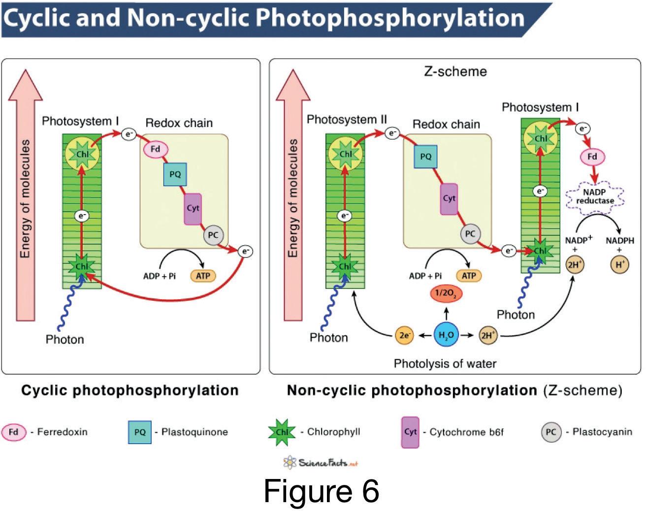
Light-independent reactions (LIR)
Light-independent reactions (LIR), also known as the Calvin cycle, occur in the stroma of the chloroplast and do not require light energy [19]. The Calvin cycle consists of three stages: carbon fixation, reduction, and regeneration [20]. In the fixation phase, the enzyme RuBisCO catalyses the combination of Ribulose Bisphosphate (RuBP) which is a five carbon compound, with a carbon atom from carbon dioxide to form a six-carbon compound [21]. However, the six-carbon compound produced is unstable; it will split into two shorter chains after its formation, each with three carbons. The chain is known as 3-phosphoglycerate (3-PGA) [23]. - Figure 7 is the Calvin cycle and Figure 8 shows the structure of 3-PGA.
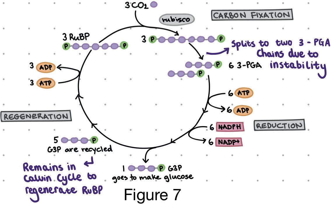
94 Scientific Harrovian 2022
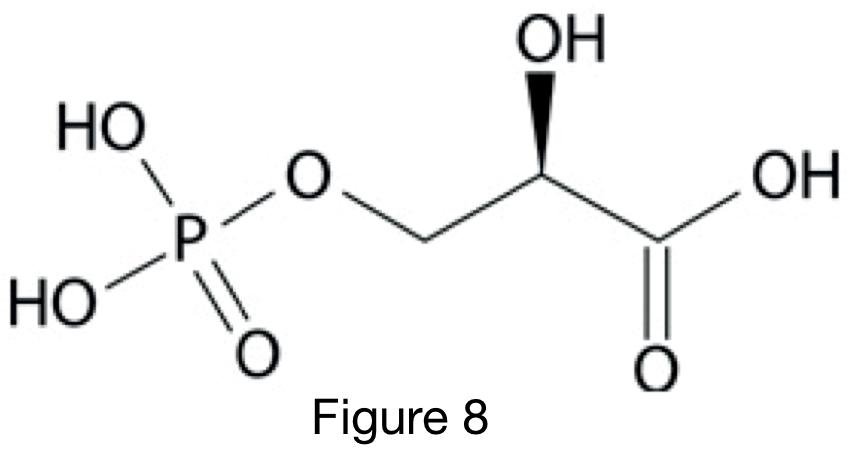
After the formation of 3-PGA, the Calvin cycle enters the reduction stage where ATP and NADPH from the light-dependent reactions (LDR) are used to convert 3-PGA into Glyceraldehyde 3 Phosphate (G3P) [21]. Both 3-PGA chains receive a phosphate molecule from ATP to form 1,3-biphosphoglycerate. This process results in the conversion of ATP into ADP. NADPH donates electrons to reduce 1,3-biphosphoglycerate, as NADPH loses electrons, it is oxidised to form NADP+. 1,3 -biphosphoglycerate also loses a phosphate group to form G3P [22].
During the regeneration process, one G3P molecule is involved in the production of glucose (C₆H₁₂O₆), while the remaining G3P molecules will remain in the Calvin cycle to regenerate RuBP. The RuBP molecules regenerated from G3P will be involved in the synthesis of glucose when more carbon dioxide enters the stomata [23].
The importance of photosynthesis is often overlooked, it is an essential and fundamental process which is continuously occurring around us. It plays a vital role in generating oxygen which we need as well as serving as a food source for numerous organisms in the food chain. Hence, it is important to understand the significance of this process and what occurs during it as this article aims to elucidate.
Bibliography
[1] Encyclopedia Britannica. (n.d.). Chloroplast. [online] Available at: https://www.britannica.com/science/chloroplast [Accessed 18 Oct. 2022].
[2] Martin, L. (2019). What Are the Roles of Chlorophyll a & b? Sciencing. Retrieved from https://sciencing.com/what-are-the-roles-of-chlorophylla-b-12526386.html
[3] Admin. (2020, September 25). Difference between chlorophyll a and chlorophyll b - byjus. BYJUS. Retrieved November 22, 2022, from https:// byjus.com/biology/difference-between-chlorophyll-a-and-chlorophyl-b/
[4] U.S. National Library of Medicine. (n.d.). Home - books - NCBI. National Center for Biotechnology Information. Retrieved November 22, 2022, from https://www.ncbi.nlm.nih.gov/books
[5] Admin. (2022). Chloroplast- diagram, Structure and function of chloroplast. BYJUS. Retrieved November 22, 2022, from https://byjus.com/biology/chloroplasts/
[6] “Light-dependent reactions. Light-Dependent Reactions - an overview | ScienceDirect Topics.” (n.d.). Retrieved November 22, 2022, from https:// www.sciencedirect.com/topics/agricultural-and-biological-sciences/light-dependent-reactions#:~:text=ATP%20and%20NADPH%20are%20energy,the%20next%20stage%20of%20photosynthesis.&text=The%20chlorophyll%20molecule%20regains%20the,dioxygen%20(O2)%20molecule.”
[7] FoodCrumbles. (2022). The Science of Color in Fruits and Vegetables. [online] Available at: https://foodcrumbles.com/colours-in-fruits-vegetables/#:~:text=In%20fruits%20and%20vegetables%20almost [Accessed 15 Dec. 2022].
[8] oakesrl1. (2010). On the Hidden Colors in Leaves: What are the Functions of Those Yellow and Orange Pigments We See in the Fall? [online] Appalachian State University Department of Biology. Available at: https://biology.appstate.edu/fall-colors/hidden-colors-leaves-what-are-functionsthose-yellow-and-orange-pigments-we-see-fall [Accessed 15 Dec. 2022].
[9] Libretexts. (2018). 8.5: The Light-Dependent Reactions of Photosynthesis - Absorption of Light. Biology LibreTexts. Retrieved December 15, 2022, from https://bio.libretexts.org/Bookshelves/Introductory_and_General_Biology/Book%3A_General_Biology_(Boundless)/08%3A_Photosynthesis/8.05%3A_The_Light-Dependent_Reactions_of_Photosynthesis_-_Absorption_of_Light
[10] Khan Academy. (2016). Light-Dependent Reactions (Photosynthesis Reaction) (Article). Retrieved December 26, 2022, from https://www.khanacademy.org/science/ap-biology/cellular-energetics/photosynthesis/a/light-dependent-reactions
[11] BYJUS. (n.d.). Difference between Photosystem 1 and Photosystem 2. [online] Available at: https://byjus.com/neet/difference-between-photosystem-1-and-photosystem-2/ [Accessed 26 Dec. 2022].
[12] NASA. (2017). Background: Atoms and Light Energy. [online] Nasa.gov. Available at: https://imagine.gsfc.nasa.gov/educators/lessons/xray_ spectra/background-atoms.html [Accessed 26 Dec. 2022].
95 Chemistry and Biology
[13] Khan Academy. (2016). Conceptual overview of light dependent reactions. [online] Available at: https://www.youtube.com/watch?v=vEsAtC9d_ MQ [Accessed 26 Jan. 2023].
[14] Khan Academy (2016b). Light-dependent reactions (photosynthesis reaction) (article). [online] Khan Academy. Available at: https://www.khanacademy.org/science/ap-biology/cellular-energetics/photosynthesis/a/light-dependent-reactions [Accessed 26 Jan. 2023].
[15] byjus.com. (n.d.). Where is ferredoxin NADP+ reductase FNR found in the leaf? [online] Available at: https://byjus.com/question-answer/ where-is-ferredoxin-nadp-reductase-fnr-found-in-the-leaf-stroma-of-thylakoidwithin-the-photosystemlumen-1/ [Accessed 26 Jan. 2023].
[16] Moolna, A. and Bowsher, C.G. (2010). The physiological importance of photosynthetic ferredoxin NADP+ oxidoreductase (FNR) isoforms in wheat. Journal of Experimental Botany, [online] 61(10), pp.2669–2681. doi:10.1093/jxb/erq101.
[17] Fowler, S., Roush, R. and Wise, J. (n.d.). Light-Dependent Reactions of Photosynthesis. https://bio.libretexts.org/Bookshelves/Introductory_ and_General_Biology/Book%3A_Concepts_in_Biology_(OpenStax)/05%3A_Photosynthesis/5.02%3A_The_Light-Dependent_Reactions_of_Photosynthesis.
[18] www.nature.com. (n.d.). ATP | Learn Science at Scitable. [online] Available at: https://www.nature.com/scitable/definition/atp-318/#:~:text=When%20one%20phosphate%20group%20is [Accessed 27 Jan. 2023].
[19] Hochmal, A.K. (2015). Light-Independent Reactions - an overview | ScienceDirect Topics. [online] www.sciencedirect.com. Available at: https:// www.sciencedirect.com/topics/biochemistry-genetics-and-molecular-biology/light-independent-reactions#:~:text=Photosynthesis%20occurs%20 in%20the%20chloroplasts. [Accessed 25 Mar. 2023].
[20] Ha, M., Morrow, M. and Algiers, K. (2020). 13.6: Light-independent Reactions. [online] Biology LibreTexts. Available at: https://bio.libretexts. org/Bookshelves/Botany/Botany_(Ha_Morrow_and_Algiers)/Unit_3%3A_Plant_Physiology_and_Regulation/13%3A_Photosynthesis/13.06%3A_ Light-independent_Reactions [Accessed 25 Mar. 2023].
[21] Symington, C. (2014). Nature’s smallest factory: The Calvin cycle - Cathy Symington. YouTube. Available at: https://www.youtube.com/ watch?v=0UzMaoaXKaM [Accessed 25 Mar. 2023].
[22] Khan academy (2021). The Calvin cycle (article) | Photosynthesis. [online] Khan Academy. Available at: https://www.khanacademy.org/science/ ap-biology/cellular-energetics/photosynthesis/a/calvin-cycle [Accessed 25 Mar. 2023].
[23] Molnar, C. and Gair, J. (2015). 5.3: The Calvin Cycle. opentextbc.ca. [online] Available at: https://opentextbc.ca/biology/chapter/5-3-the-calvin-cycle/#:~:text=One%20of%20the%20three%2Dcarbon%20PGA%20molecules%20is%20used%20to%20make%20a%20sugar%20molecule%20 while%20the%20other%20two%20PGA%20molecules%20are%20used%20to%20regenerate%20RuBP%20so%20the%20cycle%20can%20continue. [Accessed 25 Mar. 2023].
[24] Fiedor, J. and Burda, K. (2014). Potential Role of Carotenoids as Antioxidants in Human Health and Disease. Nutrients, [online] 6(2), pp.466–488. Available at: https://www.ncbi.nlm.nih.gov/pmc/articles/PMC3942711/ [Accessed 25 Mar. 2023].
[25] Mercy Education media (2019). Cyclic and Noncyclic Photophosphorylation. YouTube. Available at: https://www.youtube.com/watch?v=cdSf8SBOLwM [Accessed 26 Mar. 2023].
[26] Yahia, E. (2019). Photophosphorylation - an overview | ScienceDirect Topics. [online] www.sciencedirect.com. Available at: https://www.sciencedirect.com/topics/agricultural-and-biological-sciences/photophosphorylation#:~:text=Photophosphorylation%20is%20the%20conversion%20of [Accessed 26 Mar. 2023].
[27] Bartee, L., Shriner, W. and Creech, C. (2017). The Light-Dependent Reactions. openoregon.pressbooks.pub. [online] Available at: https://openoregon.pressbooks.pub/mhccmajorsbio/chapter/8-3-the-two-parts-of-photosynthesis-light-dependent-reactions/#:~:text=Generating%20Another%20 Energy%20Carrier%3A%20NADPH&text=As%20the%20electron%20from%20the [Accessed 26 Mar. 2023].
[28] Hashimoto, H., Uragami, C. and Cogdell, R.J. (2016). Carotenoids and Photosynthesis. Sub-cellular biochemistry, [online] 79, pp.111–39. doi:https://doi.org/10.1007/978-3-319-39126-7_4.
[29] Engelking, L.R. (2018). Electron Transport Chain - an overview | ScienceDirect Topics. [online] Sciencedirect.com. Available at: https://www. sciencedirect.com/topics/neuroscience/electron-transport-chain [Accessed 25 May 2023].
Figures used
[Figure 1] - Wikipedia Contributors (2019). Chlorophyll b. [online] Wikipedia. Available at: https://en.wikipedia.org/wiki/Chlorophyll_b [Accessed 25 Mar. 2023].
[Figure 2] - Encyclopedia Britannica (2018). chloroplast | Function, Location, & Diagram. In: Encyclopædia Britannica. [online] Available at: https:// www.britannica.com/science/chloroplast [Accessed 25 Mar. 2023].
[Figure 3] - commons.wikimedia.org. (2006). Plik:Beta-carotene.png – Wikipedia, wolna encyklopedia. [online] Available at: https://pl.m.wikipedia. org/wiki/Plik:Beta-carotene.png [Accessed 25 Mar. 2023].
[Figure 4] - Guidi, L., Tattini, M. and Landi, M. (2017). Figure 2. Chlorophyll a, b and carotenoids absorbance spectra. [online] ResearchGate. Available at: https://www.researchgate.net/figure/Chlorophyll-a-b-and-carotenoids-absorbance-spectra_fig1_317151195 [Accessed 25 Mar. 2023].
[Figure 5] - Khan Academy (2016). Light-dependent reactions (photosynthesis reaction) (article). [online] Khan Academy. Available at: https://www. khanacademy.org/science/ap-biology/cellular-energetics/photosynthesis/a/light-dependent-reactions [Accessed 25 Mar. 2023].
[Figure 6] - Dhali, Dipa. “Cyclic & Non-Cyclic Photophosphorylation: Definition & Difference.” Science Facts, 3 Sept. 2021, www.sciencefacts.net/ cyclic-and-non-cyclic-photophosphorylation.html. Accessed 24 May 2023.
[Figure 7] - Khan academy (2021). The Calvin cycle (article) | Photosynthesis. [online] Khan Academy. Available at: https://www.khanacademy.org/ science/ap-biology/cellular-energetics/photosynthesis/a/calvin-cycle [Accessed 25 Mar. 2023].
[Figure 8] - “3-Phosphoglyceric Acid.” Hyperphysics.phy-Astr.gsu.edu, hyperphysics.phy-astr.gsu.edu/hbase/Organic/3pga.html. [Accessed 23 May 2023].
96 Scientific Harrovian 2022
Synthesised MEAT
by Fabiola Chong
97 Chemistry and Biology
INTRO INTO CARBON EMISSIONS AND MEAT INDUSTRY’S RESPONSIBILITY
With 2021’s carbon emissions of 37.12 billion metric tons introducing a record high for our planet since 2010, it is safe to say we have caught ourselves amid a climate crisis- but what does this entail? From rising sea levels to the accelerated death of mankind, the consequence of a warming planet should be one we all anticipate with fear. Our planet’s atmospheric carbon dioxide (CO2) concentration was at 280 ppm at the start of the industrial revolution and has increased by a whole 50% at 421 ppm as of 2021;, the culprit behind this spur in carbon dioxide concentrations can be found, as unlikely as it seems, in plain sight between the toasted bunsfolds of your cheeseburger or on the plate that carries your slab of steak.
The animal agriculture industry is one of the biggest unspoken villains behind global carbon emissions generating 11% of our planet’s greenhouse gas emissions, with livestock farming being responsible for a whole 5.9%. Livestock farming is a section of the agriculture industry that is unique in its diversity in absolutely demolishing our planet, from deforestation to mirages of methane cow flatulence, it seems they’ve mastered pollution unlike any other industry. Due to the rising demand for meat, more so beef and red meats, an increase in deforestation has come with it. Deforestation is “the purposeful clearing of forested land” [3] to make space for agricultural production. and It is detrimental to the environment due to the masses of stored carbon dioxide found in trees released at once into the atmosphere.
DEFORESTATION AND THE ATMOSPHERE
The sun emits electromagnetic radiation (EM) that comes in waveform. Photons are the tiny particles made up of electromagnetic radiation that are responsible for warming our planet, some of which are absorbed by the Eearth’s surface or by carbon dioxide when it is re-emitted fromby the Eearth’s surface. A common belief is that carbon dioxide acts as a blanket that traps heat within the atmosphere, but, one may ask, how does the heat have a chance to permeate in the first place? This is due to the sun ray’s wavelength being much shorter than the required 15 microns [4] that carbon dioxide usually absorbs. However, when the Eearth absorbs this light and releases it back to space, the wavelength of the rays increases and there are lower frequencies of infra-red radiation, therefore allowing the carbon dioxide to trap them due to the bonds vibrating, causing the molecule to undergo symmetrical and asymmetrical stretching as well as bending. An important point to note is that no other greenhouse gas absorbs light at a wavelength of around 15 microns, so carbon dioxide is largely responsible for trapping heat and accelerating global warming[4], not to mention itsit's atmospheric lifetime of approximately 300-1000 years [5].
MEAT INDUSTRY AND METHANE GAS
Livestock within the agriculture industry is responsible for 14.5% of greenhouse gases, due to reasons regarding land usage, animal waste, enteric fermentation, and feed production. Cows are infamous for their methane production and is a result of their enteric fermentation. Enteric fermentation is when anaerobic microbes, mostly a part of the archaea domain of bacteria such as methanogens, within the digestive systems, more specifically the rumen, of ruminant animals such as cows and sheep break down fiber and carbohydrates for efficient absorption. The unfortunate byproduct of this process is methane (CH4), which is part of the greenhouse gases and negatively affects our environment. This is due to its ability being 25% more efficient in trapping heat than carbon dioxide. Although methane has a much shorter atmospheric lifetime (12 years) than carbon dioxide, and has the potential to create ozone (O3) when oxidized , methane takes an extremely long time to oxidize and the amount of methane being released into the atmosphere results in a larger net accumulation than ozone production. Methane consists of a single molecule of carbon bonded to four other hydrogen atoms, the carbon-hydrogen bond acts as a spring that is responsible for methane’s frequency. Due to the nature of the bond, methane’s absorption bands are atis at 1.7, 2.3, 3.3, and 7.6μm which means it has the capability to catch more photons at different frequencies released by earth to reflect it back. Methane naturally exists in much smaller quantities than carbon dioxide, with it only taking up 0.00017% of the atmosphere, but the demand for meat deriving from ruminant animals means the methane being produced from these animals have significantly increased the methane levels in the atmosphere.
98 Scientific Harrovian 2022
CULTURED MEAT
The option to completely eradicate the agribusiness is tempting, but we can alternatively make conscious choices in our daily lives that can reduce our carbon footprint. In terms of the agribusiness industry, this does not have to mean converting into veganism, or committing to plant based meats that (lets be honest here) tastes nothing like meat, thanks to the discovery of cultured meat.
STEM CELLS
Cultured meat is made first through the extraction of stem cells. A variety of stem cells have been considered such as embryonic stem cells, in which an embryo is extracted 3 days post fertilization to obtain a blastocyst. The inner mass of a blastocyst is used due to its pluripotency or ability to specialize into most cells such as muscle cells except for placental cells. An issue with embryonic stem cells is its inclination to instability due to its hypersensitivity towards its environment and growth substrate, making it incredibly difficult to create a stem cell line to work with. A stem cell line is a group of cells that can be continuously cultured in-vitro to use in projects including but not limited to multiple kinds of research such as stem cell research, the search for regenerative medicine, and in this case, the perfection of cultured meat. Its instability is why scientists have invented an alternative to blastocysts with the use of cellular reprogramming to create induced pluripotent stem cells (iPSC) that havehas all the benefits of a pluripotent stem cell without a blastocysts instability. A huge benefit of iPSCs is that they can be derived from almost any kind of adult somatic cells such as white blood cells with cellular reprogramming, and further specialized in a controlled manner into the muscle cells that are needed for meat culturing. How do already pre-differentiated cells such as white blood cells convert back to an embryonic-likeembryonic like state of pluripotency? The answer was broughtis brought by two scientists back in 2003, during an experiment in which 4 protein transcription factors, Oct3/4, Sox2, c-Myc, and Klf4 now known as Yamanaka factors were combined and added to mice embryonic and adult fibroblast cultures. [10] The cultures were reprogrammed into pluripotent cells and managed to maintain this state of pluripotency with the Oct3/4 and Nanog factor, and this phenomenon can now be applied to meat culturing. Another stem cell option that is frequently used is the myosatellite or satellite muscle cell, which can be obtained through a biopsy on an anaesthetized animal and is found between the sarcolemma, the plasma membrane of muscle cells, and the basal lamina, a layer that the epithelial cells sit on and produce. Satellite cells are multipotent, which means they can develop into more than one type of cell but are much more limited compared to the iPSCs. An issue with satellite cells that is not found with iPSCs is its invasive nature towards animals when obtaining them, since these cells are usually found in bone marrow or close to the spinal cord, and can at times, require the death of the animal. This makes the purpose of meat culturing completely redundant.
The satellite cells are allowed to differentiate into myocytes and proliferate, which will result in the formation of muscle fibers after they merge, and the iPSCs are left to proliferate post specialization by submerging the respective cell lines into culture media in a bioreactor, which contains amino acids, several macro and micronutrients, vitamins and minerals, but most importantly, fetal bovine serum(FBS). Fetal bovine serum is used universally across a majority of cellular research that requires in vitro cell culture as an important supplement due to its ability to be used with almost all types of cells. However, major ethical issues surround the use of fetal bovine serum, as it is obtained through the slaughter of cow fetuses immediately after birth by cardiac puncture and drainagedraining of their blood. The entire process is done without the use of anesthesia to maintain purity and is a slow, painful death. The blood is then refined and used as fetal bovine serum. Not to mention, it costs 2000 USD for a litre of FBS to be produced, and is expected to be constantly changed out during the culturing of cells for meat due to the myocytes-turned-muscle fibers constantly contracting post differentiation, producing lactic acid and changing the PH of the medium, making it an unsuitable environment for survival or growth. Due to the controversy and high costs surrounding the production of fetal bovine serum, alternatives to the serum have been sought and have been successful with cell growth, with some alternatives completely eliminating the use of
99 Chemistry and Biology
serum completely. The National University of Singapore recently discovered an unconventional method of cell culturing with the use of magnetism which they believe to be a greener approach in contrast with the alternatives of excessive drug usage and genetically modifying the organisms to achieve cell growth. They do so by using a pulsed magnetic field to stimulate the release of certain molecules referred to as ‘muscle secretomes’ that are integral to the growth and development of these cells. Although this technology is relatively new, it shows great promise in the elimination of FBS in future production of cultured meat.

Source: https://news.nus.edu.sg/novel-technique-to-grow-meat-in-the-lab-using-magnetic-field/
SCAFFOLDING
EXTRACELLULAR MATRIX
Scaffolding is done within the bioreactor and is responsible for the outcome of structure, flavor, and texture of the meat by guiding cell differentiation. A bioreactor is kept at a constant temperature of 37 degrees to allow the cell’s enzymes to operate at its optimum and prevent denaturing. Sterilisation is crucial in meat culturing, as any changes may lead to an infected batch of cells and must be completely thrown out, which is why bioreactors tend to be made out of steel or glass with a disposable for sterilization liner to be carried out much easily. Scaffolding technologies are an area of research typically found within tissue engineering and regenerative medicine, in which edible scaffolds are used to allow the cells to bind onto and form the foundations of its 3D structure. The extracellular environment the cell is in is integral to the specialization of the cell, with the extracellular matrix (ECM) being a huge contributor to the environment. The ECM is composedcomprised of collagen that is connected to integrin by fibronectin and is embedded into the cell’s phospholipid bilayer. Due to early embryos essentially being made up of stem cells, they will undergo embryogenesis and cell specialization that is, unsurprisingly, controlled by their extracellular environment. Integrin is responsible for a process known as mechanotransduction, where the integrins essentially take the role of mechanosensors that mediate downstream effector proteins creating focal adhesion complexes between the cytoskeleton of the cell to the matrix. These connections then affect the cell’s response to their extracellular environment which will ultimately result in itsit’s polarity, migration and differentiation into muscle cells. Other factors such as ECM densities, gradients and 3D topography are also responsible for cellular response and outcome. All of these components of the cell are taken into consideration during the scaffolding process which truly reveals the tedious nature of cell culturing due to the cell’s sensitivity to its environment, which is why microcarriers are also used to ensure the cells survive their proliferation stage when dropped into an environment completely foreign to their in vivo origins as it can potentially lead to anoikis in which […]. This is usually because the cells are unable to undergo suspension growth or grow as spheroids, which is where the use of microcarriers comescome in. The microcarriers are essentially there to provide a large surface area to volume ratio to encourage high densities of cells to form and expand. The edible scaffolds tend to be made out of gelatin, and are usedis
100 Scientific Harrovian 2022
used to create that thick and structured organic meat effect. This is done by the scaffold controlling the attachment, maturation and differentiation of cells by considering the scaffold’s porosity and mechanical properties. The biomaterials the scaffold is made of is also extremely important, as each material will bring about its downstream properties. These materials can range from algae to spider silk, and are all unique in the properties they bring to the scaffold, and therefore structure of the meat. An issue with scaffolds is the biomaterials deriving from animal tissues, which some may argue completely makes the purpose of lab grown meat obsolete if it means farming animals for gelatin, which is why alginate or chitosan based scaffolds are considered. However, their functionality is severely limited due to them lacking the motifs: Arg- Gly-Asp and Ile-Lys-Val-Ala-Val that promote cell adhesion.
HARVESTING AND PROCESSING
Once enough cells have been harvested, the scaffolds are separated and the meat is processed. The meat patty is the easiest type of meat product to be made due to its easy assembly, hence the first successful cultured meat product being a hamburger patty in 2013. Steaks on the other hand, are much more difficult to grow due to theirits thickness and susceptibility to cell death as a result. A huge benefit of cultured meat besides its low ecological impact is its production time. A conventional chicken can take 7-12 weeks to grow from fertilization of the egg to slaughter, with a cow taking a whole 112 weeks. With cultured meats, disregarding species, the time taken to grow from entering the bioreactors to harvest is only at 5-7 weeks and requires significantly less resources. Culturing meat on an industrial level will require 89% less water, 99% less land usage compared to traditional livestock farming and will potentially be able to lower greenhouse gas emissions by 96% [18]. With FDA’s approval of cultured meat being safe to eat, Singapore has been the first country to approve the sale of lab grown meat in December 2020, and other countries have also been considering its sale, with the USA being responsible for 60% of global investments on cultivated meat. When cultured meat hits the global market and COMPLETELY alters your life, will you take a chance on it?
101 Chemistry and Biology
Citations:
[1] Tiseo, I. (2023, February 6). Annual global emissions of carbon dioxide 1940-2021. Google search. Retrieved March 23, 2023, from https:// www.google.com/search?q=Global%2BCO2%2Bemissions%2Bby%2Byear&sa=X&ved=2ahUKEwiU_5-0kfH9AhVwTGwGHXysCvAQ1QJ6BAhSEAE&biw=1440&bih=789&dpr=2&safe=active&ssui=on
[2] Stein, T. (n.d.). Carbon dioxide now more than 50% higher than pre-industrial levels. National Oceanic and Atmospheric Administration. Retrieved March 23, 2023, from https://www.noaa.gov/news-release/carbon-dioxide-now-more-than-50-higher-than-pre-industrial-levels
[3] “Deforestation.” Education, https://education.nationalgeographic.org/resource/deforestation/.
[4] “How Do Greenhouse Gases Trap Heat in the Atmosphere?” MIT Climate Portal, 19 Feb. 2021, https://climate.mit.edu/ask-mit/how-dogreenhouse-gases-trap-heat-atmosphere.
[5] Buis, Alan. “The Atmosphere: Getting a Handle on Carbon Dioxide – Climate Change: Vital Signs of the Planet.” NASA, NASA, 16 Nov. 2022, https://climate.nasa.gov/news/2915/the-atmosphere-getting-a-handle-on-carbon-dioxide/#:~:text=Carbon%20dioxide%20is%20a%20 different,timescale%20of%20many%20human%20lives.
[6]“Global Greenhouse Gas Emissions Data.” EPA, Environmental Protection Agency, 15 Feb. 2023, https://www.epa.gov/ghgemissions/global-greenhouse-gas-emissions-data.
Ritchie, Hannah, et al. “Meat and Dairy Production.” Our World in Data, Our World in Data, 25 Aug. 2017, https://ourworldindata.org/ meat-production#:~:text=Global%20demand%20for%20meat%20is,agricultural%20land%20and%20freshwater%20use.
“Answers to Your Questions about Stem Cell Research.” Mayo Clinic, Mayo Foundation for Medical Education and Research, 19 Mar. 2022, https://www.mayoclinic.org/tests-procedures/bone-marrow-transplant/in-depth/stem-cells/art-20048117.
the United Nations, Food and Agriculture Organisation of. “Livestock Solutions for Climate Change - Food and Agriculture Organization.” Livestock Solutions for Climate Change, 2017, https://www.fao.org/3/i8098e/i8098e.pdf.
AgLEDX. “Enteric Fermentation: Emissions & Mitigation Options " Agled.” AgLEDx Resource Platform, 17 Nov. 2022, https://agledx.ccafs.cgiar. org/emissions-led-options/sources-sinks/enteric-fermentation/#:~:text=Enteric%20fermentation%20occurs%20when%20anaerobic,otherwise%20 would%20not%20be%20digestible.
CJ;, Morgavi DP;Forano E;Martin C;Newbold. “Microbial Ecosystem and Methanogenesis in Ruminants.” Animal : an International Journal of Animal Bioscience, U.S. National Library of Medicine, 2010, https://pubmed.ncbi.nlm.nih.gov/22444607/#:~:text=Methanogenesis%20is%20 performed%20by%20methanogenic,functional%20niche%20in%20the%20ecosystem.
Environmental Protection Agency, United States. “Importance of Methane.” EPA, Environmental Protection Agency, 2022, https://www.epa.gov/ gmi/importance-methane#:~:text=Methane%20is%20more%20than%2025,due%20to%20human%2Drelated%20activities.
Libretexts, Chemistry. “Methane and Global Warming.” Chemistry LibreTexts, Libretexts, 11 Mar. 2023, https://chem.libretexts.org/Ancillary_ Materials/Exemplars_and_Case_Studies/Exemplars/Environmental_and_Green_chemistry/Methane_and_Global_Warming.
Byrom, R.E, and K.P Shine. “Year‐2020 Global Distribution and Pathways of Reservoir Methane and ...” AGU Pubs, 2022, https://agupubs. onlinelibrary.wiley.com/doi/10.1029/2020GB006888.
Frazier, Reid. “Why Methane Is Such a Potent Greenhouse Gas.” The Allegheny Front, 14 May 2020, https://www.alleghenyfront.org/why-methane-is-such-a-potent-greenhouse-gas/.
“Cultivated Meat Cell Lines: Deep Dive: GFI.” The Good Food Institute, 15 Oct. 2021, https://gfi.org/science/the-science-of-cultivated-meat/deepdive-cultivated-meat-cell-lines/.
[10]Takahashi, Kazutoshi, and Shinya Yamanaka. “Induction of Pluripotent Stem Cells from Mouse Embryonic and Adult Fibroblast Cultures by Defined Factors.” Cell, vol. 126, no. 4, 2006, pp. 663–676., https://doi.org/10.1016/j.cell.2006.07.024.
Verma, Rajneesh, et al. “IPSC Technology: An Innovative Tool for Developing Clean Meat, Livestock, and Frozen Ark.” Animals, vol. 12, no. 22, 2022, p. 3187., https://doi.org/10.3390/ani12223187.
Snijders, Tim, et al. “Satellite Cells in Human Skeletal Muscle Plasticity.” Frontiers, Frontiers, 23 Sept. 2015, https://www.frontiersin.org/articles/10.3389/fphys.2015.00283/full#:~:text=The%20term%20%E2%80%9Csatellite%20cell%E2%80%9D%20was,%2C%20proliferate%20 and%2For%20differentiate.
“What Is the Difference between Totipotent, Pluripotent, and Multipotent?” What Is the Difference between Totipotent, Pluripotent, and Multipotent? | NYSTEM, https://stemcell.ny.gov/faqs/what-difference-between-totipotent-pluripotent-and-multipotent#:~:text=Multipotent%20 cells%20can%20develop%20into,stem%20cells%20are%20considered%20multipotent.
Jochems CE;van der Valk JB;Stafleu FR;Baumans V; “The Use of Fetal Bovine Serum: Ethical or Scientific Problem?” Alternatives to Laboratory Animals : ATLA, U.S. National Library of Medicine, 2002, https://pubmed.ncbi.nlm.nih.gov/11971757/#:~:text=Abstract,without%20any%20 form%20of%20anaesthesia.
Singapore, National University of. “How Magnets Could Improve Cell-Based Meat.” Futurity, 18 Oct. 2022, https://www.futurity.org/cell-basedmeat-magnets-2816162-2/.
Singapore, National University of. NUS Scientists Develop Novel Technique to Grow Meat in the Lab Using Magnetic Field, National University of Singapore, 1 Nov. 2022, https://news.nus.edu.sg/novel-technique-to-grow-meat-in-the-lab-using-magnetic-field/. Institute, Good Food. “Cultivated Meat Scaffolding: Deep Dive: GFI.” The Good Food Institute, 6 Aug. 2021, https://gfi.org/science/the-science-of-cultivated-meat/deep-dive-cultivated-meat-scaffolding/.
[18]Reiss, Jacob, et al. “Cell Sources for Cultivated Meat: Applications and Considerations throughout the Production Workflow.” International Journal of Molecular Sciences, U.S. National Library of Medicine, 13 July 2021, https://www.ncbi.nlm.nih.gov/pmc/articles/PMC8307620/.
102 Scientific Harrovian 2022
artificial PHOTOSYNTHESIS
to achieve carbon neutrality
 by Bess Chau
103 Chemistry and Biology
Synopsis
The goal of this article is to uncover the qualities of plants that deviate from normal limiting factors and combine them all through genetic modification into the ‘perfect plant’ that can photosynthesise for as long as possible, thus be able to take in more carbon dioxide emissions and achieve carbon neutrality.
The problem with carbon emissions
Ever since the industrial revolution, the rate of carbon emissions has been increasing at an alarming speed. The development of technologies and the strive for efficiency and money has caused a vicious cycle that has caused carbon emissions to rise.
The correlation between carbon emissions and damage to both us and our earth exists in 2 ways :
1. Direct influence regarding the greenhouse effect
Carbon particles react with oxygen to form carbon dioxide, a common greenhouse gas which absorbs the heat radiated from the sun and radiates it in all different directions, including back to our planet. This causes a rise in the earth’s temperatures and thus contributes to global warming.
Several detrimental effects of Global warming include the melting of the ice caps, seasonal temperature extremes, heavy rainfall and changing habitat ranges for plants. Judging the size of the oceans and lands of the earth, it would take an abundance of heat energy to raise the yearly temperature of the earth, but data collected from scientists that measure temperatures all across the globe (undergoing the measurements of the difference between observed temperature and the long-term average temperature in different locations) collectively show an upward trend in carbon emission rates, signifying the severity of human activity. [1]
2. The depletion of the ozone layer (also known as the thinning of the ozone layer)
Note, the depletion of the ozone layer isn’t the main cause of climate change. Whilst it does factor in global warming, it is minimal in comparison to human activity which is a main factor since it directly releases carbon emissions. However, the depletion of the ozone layer has a different effect on us as data shows a direct correlation in the rise in skin cancer with increased exposure of the UV rays from the sun, correlation increasing with the thinning of the protective ozone layer. [2]
The ozone layer is extremely important since the ozone particles collect and bunch in the upper stratosphere, where it acts as a shield against harmful portions of the sun’s radiation, especially the UV-B part of the electromagnetic spectrum. It also prevents heat in greenhouse gases from being absorbed and reflected back onto earth, heating up the core. [3]
The depletion of the ozone layer is mainly caused by the CFC gas since CFCs break down ozone molecules within the layer. Whilst carbon particles don’t break down ozone molecules themselves, the rise of carbon emissions causes a cooling within the stratosphere, which (1) makes the ozone layer more vulnerable as it increases in sensitivity, and (2) form stratospheric clouds which contributes to ozone loss when many clouds form. [4]
Fortunately for us, the ozone layer is expected to recover and thicken again, due to CFCs being banned in many countries. The UN Education Program predicts that maintaining current activity could almost guarantee the recovery of the ozone layer by 2066 over Antarctica, 2045 over the Arctic and 2040 for the rest of the world. However, to prevent the slowing of its recovery, we must do our best to from ex-
104 Scientific Harrovian 2022
acerbating current damages such as the ‘ozone hole’ or extra problems in the future (this means actively preventing all types of activities that could slow the pace of ozone recovery, including stopping the rise in carbon emissions)
The solution to carbon emissions - photosynthesis
Understanding the severity of carbon emissions, we need to find a solution which can not only limit the carbon emissions, but also reduce the carbon dioxide within the atmosphere. A solution discussed in today’s article will be, specifically photosynthesis.
Photosynthesis is a process that allows carbon dioxide to be taken in and oxygen to be released through gas exchange. Therefore, plants and humans are able to form a symbiotic relationship through photosynthesis since plants take in carbon dioxide that we exhale and produce oxygen for us to inhale.
Limiting factors of photosynthesis
One underlying problem of photosynthesis, however, are the limiting factors that decrease the rate of photosynthesis.
For instance:
1. Temperature (extreme weather conditions)
2. Salinity
3. Insufficient sunlight
4. Insufficient water
5. Bacteria
1. Temperature:
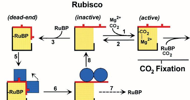

The process of photosynthesis is an enzyme-driven chemical reaction that provides kinetic energy towards Rubisco.(Rubisco or Ribulose-1,5-bisphosphate carboxylase/oxygenase are the enzymes within photosynthesis. They form an enzyme substrate complex with CO2 to give rise to photorespiration [5] [6])
When the temperature is too cold, the enzymes that react with substrates to form enzyme-substrate complexes may not have enough kinetic energy to function. Whereas if the temperature is too hot and surpasses the optimum temperature, they melt and alter the shape of the tertiary structure of the enzymes, altering their active site, which means that enzyme-substrate complexes can no longer form and photosynthesis cannot occur. Additionally, denaturation is irreversible and the enzyme is damaged permanently.
2. Salinity:
Exposure to too much salinity can lead to the closure of the stomata(leaf pores that allow the taking in of carbon dioxide as well as the loss of water vapour for transpiration), reducing the chlorophyll content within chloroplasts or preventing plants from taking up water, inhibiting photosynthesis. Salt induced osmotic effects also adversely affect the activities of a number of stomatal enzymes involved in carbon dioxide reduction. [8]
Sometimes, high concentrations of salt in the soil could limit the water in the uptake within the roots, Sodium and Chlorine can further introduce harmful toxicities if there is a high concentration within the soil. All of these could lead to the damage of plants. [9]
105 Chemistry and Biology
3. Insufficient sunlight:
Sunlight is usually absorbed by the chlorophyll within chloroplasts, which is converted into chemical energy. The energy of the sun is used to convert water and carbon dioxide into a sugar called glucose which releases energy in the form of ATP (adenosine triphosphate); glucose can either be used in the making of cellulose or the storage of starch, for photosynthesis. Insufficient sunlight means less energy is absorbed by the plant to create glucose for food, cell-wall building and storage. .
4. Insufficient water:
Water is highly needed for all living beings. Water is taken in by plants and is oxidised (loses electrons) to form oxygen but also contributes the electrons that bond with hydrogen to the carbon atoms to form glucose. Water is also used at the most basic level to hydrate the plant and ensure that the cells within the plant don’t plasmolyse and ultimately die.
5. Bacteria:
Bacteria or virus-infected plants may cause damage to plant cells or leaves. Pathogens can cause necrosis by secreting toxins into the leaves by invading the plant tissue and multiplying within. They have a high speed of invasions and it’s even more dangerous with an open wound in a plant. Some symptoms are leaf spots(photo1), stem blights (photo with blue backdrop) or crankers (photo 3). [10.1]
Furthermore, eurythermal organisms tend to have an overall lower infection rate since bacteria cannot withstand the high temperatures and will denature. [10.2]


How to overcome these boundaries
Currently, plants showcase potential to overcome said limiting factors. They are bred from their environmental factors and developed resistance towards limiting factors through natural selection. The unique traits of different plants inspire more changes towards the plant.
Examples could include:
Euryhaline organisms


Halophiles are predominant organisms that live in salt lakes or evaporating sea water or anywhere with a salt concentration at least five times greater than that of the ocean.
- They tend to exclude salt from the cytoplasm to tolerate high concentrations of salt [11]
- Furthermore, they tolerate high concentrations of salt by maintaining high levels of water within them. It’s either by :
a) accumulating osmoprotectants : [11] Osmoprotectants : defined as - highly hydrophilic organic molecules that accumulate in cells in response to desiccation and other abiotic stresses. proteome)
106 Scientific Harrovian 2022
b) Ensuring that all enzymes and cell components adapted to the presence of high salt concentrations (more acidic amino acids than hydrophobic ones) [12]
Eurythermal organisms : organisms that tolerate a large range of temperatures
Examples include: Psychrophilic Organisms and Archaea, fungi, algae, bacteria (extreme cold)
In these organisms, their enzymes are specially adapted to still function even with lower temperatures, it comes from the proteins they produce → antifreeze proteins/ice-binding proteins which bind to and control ice crystal growth by lowering the freezing point. In addition, EPS (Extracellular polymeric substances) can trap water, nutrients and metal ions and facilitate surface adhesion(the joining of dissimilar molecules).
The organisms also undergo cellular aggregation and biofilm formation to protect enzymes against cold denaturation and autolysis.(autolysis : the breakdown of a cell and dissolving of its cell membrane, causing inside contents to digest external surrounding tissue.) They have an optimum temperature of 10-15 Degrees, lower than the average optimum temperature of a plant - which is 20-25 degrees and are capable of surviving at lower temperatures than most plants. [14]
Cactus (plants that can withstand insufficient water conditions)
Understanding cacti, they have different methods of conserving as much water as possible. Cactus are stationed in dry lands, common in the desert. Despite the cool nights, the cactus needs to conserve water in order to stay hydrated and photosynthesise. [15]
Cacti also have no leaves to prevent evaporation of water, instead it contains thick stems with lots of water vessels (xylem) to store water.
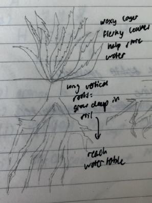
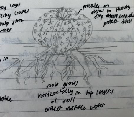
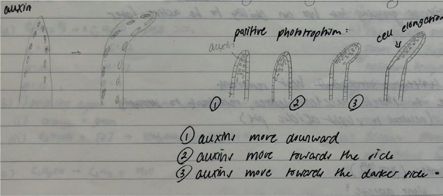
Above are 3 types of cacti and their water storage methods:


1. The first types are long vertical roots that reach water deep underground at the bottom of the soil
2. The second type are long roots that stretch horizontally to maximise collected water and nutrients in the surface level
3. The third types are large water tubers/bulbs underneath the soil
Other features contain : prickles to prevent animals from drinking water / sucrose or a waxy cuticle to help store more water and prevent water evaporation
Sunflower(they face in the direction of the sun throughout the day) + auxins
Sunflowers have the plant hormone Auxins (a breakdown of glycosides and are made of amino acids) which are produced at the tip of the stem and accumulate in the shaded area where the sun doesn’t face/ shine in. It enables the sunflower to have positive phototropism and maximise photosynthesis.
107 Chemistry and Biology
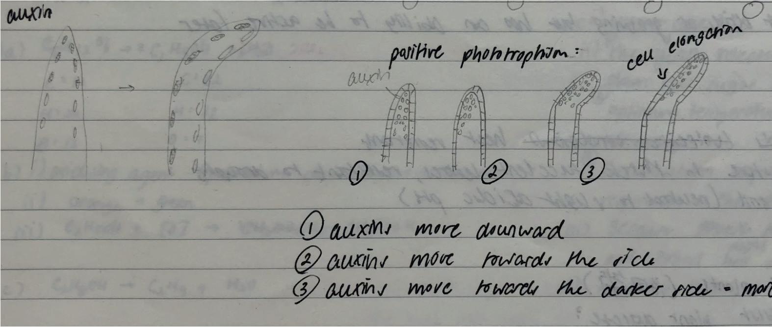

By combining all solutions into the creation of multiple plants to artificially maximise photosynthesis at a continuous rate, it is possible to guarantee large intake of carbon dioxide.
These plants have withstood factors that normally limit photosynthesis, by analysing how the plants work the way they do, combing the characteristics and providing good conditions for them to thrive in, they are the solution towards maximising photosynthesis constantly and taking in higher levels of carbon dioxide.
How to combat any diseases - a study into plants’ immune systems
Another layer of fact to incorporate is how plants deal with diseases.
The ultimate goal of defense mode is to get proteins that will help defend the plant against the invading pathogen, but there are a couple of ways to get more defensive proteins. To get a defensive protein, you have a defensive protein gene that gets copied and then that copy gets read into protein. If you want more protein you can either increase the number of copies you make or increase the speed or number of times that you read that copy.
Basic plant structure for defense
Defense structure of a plant : waxy cuticle; sharp pricks, needles, barbs; lignin on bark of a tree(impermeable for insects, hard to chew through)
The plants have built in defence structures already :
- Cactus developed sharp pricks to prevent animals from stealing its water storage
- The waxy cuticles are to keep the leaves safe from evaporation and external water/sucrose storage from entering the plant
- The lignin of a bark makes it impermeable for insects to chew through
Gene for Gene Model
The gene for gene relationship by Harold Flor states that each gene controlling resistance within the host has a corresponding specific gene within the pathogen itself that controls avirulence (ensuring the pathogenic gene doesn’t spread). This ensures that the resistance of the plant and the pathogen’s ability to spread diseases/cause diseases are both controlled by a matching pair of genes.
The genes are actually sensors that detect microorganisms entering the plant and initiate defensive mode. [16]
The plant pathogens that contain Avirulence gene induce a resistance reaction and generate a response from the plant. However, this only works when the plant has a resistance gene. The avirulence gene by the pathogen matches with the resistance gene within the plant, it can trigger primary proteins to interact
108 Scientific Harrovian 2022
with the plant’s immune system further increasing knowledge of the molecular interactions for greater disease control of plant pathogens.
The plant’s resistant gene evolves with the avirulent gene, keeping the immunity up to date. However, in cases of the avirulence gene being recessive, the gene would be able to bypass plant resistance mechanisms and cause diseases.
Methods of implementing the genes into the artificial plant
As stated before, the artificial model of the plant should be a combination of all characteristics above. A method of combining these genes is through genetic engineering or genetic modification.
There are 2 methods of genetic engineering into a plant in order to create a transgenic plant(plant that contains a gene artificially inserted)!
2 ways to create transgenic plants
1. Brute force strategy

Gene gun - firing DNA coated gold-particles at the plant cell : [17]


Through electroporation, the gene gun delivers plasmid DNA coated with gold particles into tissues into the stem of a plant. This pulses an electric field to induce transmembrane potential(the membrane of molecules to be more permeable) plant stems’ exposure to extracellular molecules. Gene guns are designed to penetrate and overcome tough cell barriers and transfect cells deep into tissues of targeted organisms. [18]
However there is still a potential for significant damage towards the tissue : it could result in cell death and loss of cell homeostasis (controlling of internal conditions to prevent the denaturing of enzymes and death of cells) and it needs to be precise aiming towards the nucleus of cells
2. Genetic Modification
Firstly, genetic modification is a process of implementing desired characteristics into the plant. The plant’s restriction enzyme cuts the plasmid of the target plant tissue (target plant tissue : the plant we are trying to genetically modify), then as the new DNA is inserted into the plasmid, it is sewn/stuck together by a ligase enzyme, forming a new transgenic plasmid. (transgenic plasmid : plasmid that is now genetically modified)
Structure of the transgenic plasmid
The usage and development of genetic modification can combine with natural selection to create a plant that can create 2 desired effects.
1. It can immediately take into effect and an abundance can help photosynthesise to the largest extent and restore some levels of carbon emissions, which is the intention of the plant
2. The second effect is the effect on nature. Hopefully these characteristics can combine and develop to form generations of different combinations within plants, spreading characteristics that can overcome limiting factors and ultimately subtly increasing the rate of carbon intake and balancing out the emissions from human activities.

Promoter - the start of the sequence for transcription
ORI - origin of replication
Antibiotic-resistant gene to the plasmid - so if the bacteria is met with the antibiotic, only cells with the plasmid will grow
Chemistry and Biology
109
Conclusion :
Overall, we hope to combine the characteristics that can help continue photosynthesis as long and effectively as possible. The construction of this ‘theoretically perfect plant’ can help take in more carbon dioxide and achieve carbon neutrality in both the long and short term.
Citations :
1. “Climate Change: Global Temperature | NOAA Climate.gov.” Www.climate.gov, www.climate.gov/news-features/understanding-climate/climate-change-global-temperature#:~:text=That%20extra%20heat%20is%20driving.
2. “Difference between Ozone Depletion and Global Warming.” Compare the Difference between Similar Terms, www.differencebetween.com/ difference-between-ozone-depletion-and-global-warming/#:~:text=The%20key%20difference%20between%20ozone. Accessed 18 Apr. 2023.
3. Hanley, Steve. “UN Report Says Ozone Layer Is Getting Thicker.” CleanTechnica, 10 Jan. 2023, cleantechnica.com/2023/01/10/un-report-saysozone-layer-is-getting-thicker/#:~:text=The%20cause%20was%20traced%20to. Accessed 18 Apr. 2023.
4. “NASA GISS: Research Features: Ozone and Climate Change.” Www.giss.nasa.gov, www.giss.nasa.gov/research/features/200402_tango/#:~:text=Ozone%20chemistry%20is%20very%20sensitive.
5. “Key Carbon Fixation Enzyme, Rubisco, Also Is Important for Sulfur Metabolism�.” The Journal of Biological Chemistry, vol. 290, no. 52, 25 Dec. 2015, p. 30669, www.ncbi.nlm.nih.gov/pmc/articles/PMC4692198/, https://doi.org/10.1074/jbc.P115.691295.
6. Jensen, R. G. “Activation of Rubisco Regulates Photosynthesis at High Temperature and CO2.” Proceedings of the National Academy of Sciences, vol. 97, no. 24, 21 Nov. 2000, pp. 12937–12938, www.pnas.org/content/97/24/12937, https://doi.org/10.1073/pnas.97.24.12937.
7. Markgraf, Bert. “What Provides Electrons for the Light Reactions?” Sciencing, 2018, sciencing.com/what-provides-electrons-for-the-light-reactions-13710477.html.
8. Hnilickova, Helena, et al. “Salinity Stress Affects Photosynthesis, Malondialdehyde Formation, and Proline Content in Portulaca Oleracea L.” Plants, vol. 10, no. 5, 22 Apr. 2021, p. 845, https://doi.org/10.3390/plants10050845.
9. Bernstein, Nirit. “Chapter 5 - Plants and Salt: Plant Response and Adaptations to Salinity.” ScienceDirect, Academic Press, 1 Jan. 2019, www. sciencedirect.com/science/article/abs/pii/B9780128127421000052.
10. “Plant Disease - Symptoms and Signs.” Encyclopedia Britannica, www.britannica.com/science/plant-disease/Symptoms-and-signs.
11. “8.15B: Extremely Halophilic Archaea.” Biology LibreTexts, 23 June 2017, bio.libretexts.org/Bookshelves/Microbiology/Microbiology_ (Boundless)/08%3A_Microbial_Evolution_Phylogeny_and_Diversity/8.15%3A_Euryarchaeota/8.15B%3A_Extremely_Halophilic_Archaea.
12. Oren, Aharon. “Bioenergetic Aspects of Halophilism.” Microbiology and Molecular Biology Reviews, vol. 63, no. 2, June 1999, pp. 334–348, https://doi.org/10.1128/mmbr.63.2.334-348.1999.
13. “Repeated Genetic Targets of Natural Selection Underlying Adaptation of Euryhaline Fishes to Changing Salinity | U.S. Geological Survey.” Www.usgs.gov, www.usgs.gov/publications/repeated-genetic-targets-natural-selection-underlying-adaptation-euryhaline-fishes. Accessed 20 Apr. 2023.
14. De Maayer, Pieter, et al. “Some like It Cold: Understanding the Survival Strategies of Psychrophiles.” EMBO Reports, vol. 15, no. 5, 26 Mar. 2014, pp. 508–517, www.ncbi.nlm.nih.gov/pmc/articles/PMC4210084/, https://doi.org/10.1002/embr.201338170.
15. “Cacti.” National Wildlife Federation, www.nwf.org/Educational-Resources/Wildlife-Guide/Plants-and-Fungi/Cacti#:~:text=To%20prevent%20water%20loss%2C%20cacti. Accessed 20 Apr. 2023.
16. The Gene-For-Gene Concept: A Central Tenet in Plant Pathology – Plants Rule. www.plantsrule.com/the-gene-for-gene-concept-a-central-tenet-in-plant-pathology/. Accessed 23 Apr. 2023.
17. “Gene Gun - an Overview | ScienceDirect Topics.” Www.sciencedirect.com, www.sciencedirect.com/topics/biochemistry-genetics-and-molecular-biology/gene-gun#:~:text=Gene%20gun%20is%20used%20to.
18. Belyantseva, Inna A. “Helios(®) Gene Gun-Mediated Transfection of the Inner Ear Sensory Epithelium: Recent Updates.” Methods in Molecular Biology (Clifton, N.J.), vol. 1427, 2016, pp. 3–26, pubmed.ncbi.nlm.nih.gov/27259918/, https://doi.org/10.1007/978-1-4939-3615-1_1.
110 Scientific Harrovian 2022


The technological and social issues that surgical robots faced and the current solution
by Elaine Zhang


111 Chemistry and Biology
As the robotic system develops, individuals continue to work on developing technology that can take the place of manual labour. Examples include the disinfection robots you frequently see in public spaces as well as sweeping robots and dishwashers. The most popular and modern robots in the surgical field, however, are typically instruments that serve to increase the precision and effectiveness of an operation. Since this robot is more akin to a doctor’s rack, he can improve their abilities.
Da Vinci robots must be mentioned when discussing surgical robots because they are the most well-known and frequently used robot. In 2022, 74.9% of Endoscopic surgeries in China were done by Da Vinci robots, performing 10 million surgeries so far. Other robots like Senhance, Micro Hand S, Versius, etc, are also focused on improving surgical precision and efficiency [1].
The first recorded use of surgical robots was in 1985 with the assistant of ‘PUMA 560 robotic surgical arm’ in a brain biopsy procedure. In 1999, Davinci robots received approval from the FDA for use in the US. This is also the first approval from FDA to a surgical robot and ‘since the first FDA clearance in 2000, the da Vinci system remained the main robotic surgical system for over 20 years’ [2].
Advantages
There are three main advantages of incorporating surgical robots in surgery. First of all, shivering from exhaustion is unavoidable especially when doctors have performed an eight-hour surgery. Hence, this is where our technology can help to minimise the effect of shivering and provide more stable ‘hands’ for the surgeon. Refer to Picture 1. It is a snapshot from 2010, from Davinci Robot’s video film, showing how precisely they can perform stitching on a grape skin(https://www.youtube.com/ watch?v=KNHgeykDXFw&t=40s)
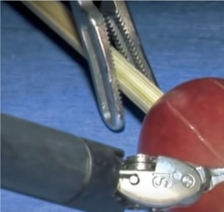
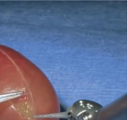

. Moreover, the precision of this equipment can also minimise the wound left on the patient after the surgery, robotic surgical systems can use the smallest incision to see the patient’s internal structure clearly [3].
However, they all have a lot of room for improvement and a lot of difficulties to solve. In this paper, I am going to introduce the challenges they faced in two major areas, external and technological.
Technological Issues
Most of the robotic surgical systems are a combination of a surgeon console, a patient side chart with robotic arms, and a 3D vision system. This precise technology provides them with a greater range of motion. This is also why most of the surgeries that involve robots are small and minimally invasive since this is where our surgery robots can show their maximum potential.
Remote surgery
The robotic surgical system’s prospects in the medical sector are promising. Through robotic surgical systems, remote surgery can become commonplace, enabling the best surgeons to perform surgeries remotely on patients in a different cities or countries. To test the limits of remote surgery, NASA proposed to perform surgery on astronauts using remote-controlled robots. As early as 2006, Surgeon Mehran Avari successfully repaired a scar on a patient who was in Aquarius underwater base through a robot, while he was on land[9]. Additionally, the 5G technologies also enabled ‘safe and efficient complex surgical procedures using telementoring with telestration in real-time, with a very high degree of surgical team satisfaction’.[10]
112 Scientific Harrovian 2022
Recognition
Most of the time in a surgery, surgeons use their knowledge in combination with both haptic and visual feedback to analyse the condition of the patient. Therefore, interaction is extremely important in a surgery. For example, if the feedback is not accurate, the surgeon could be misled to apply too much force to the suture, causing an injury or applying too little force, causing a subsequent issue [5]. Even though 3D vision systems have already provided a clear view of the operating area, it is still difficult to give the exact haptic feedback from the robotic arms to the surgeon console. Currently, there are several developing ideas to solve this problem.
Haptic feedback gloves

One of the ideas is the haptic feedback glove. It is thought that the glove would enable the doctor ‘to control robots remotely with a degree of precision approaching that of real human hands.[6]’ The design from Zhu Minglu, a researcher from national university of Singapore, and his team, uses epoxy to bond the as-fabricated sensors in each finger case and ‘the palm sensor at the centre is applied to enable the detection of normal force and shear force when in contact with an external object’[7]. The gloves also feature many microsensors and a machine learning system.Convolutional neural network is a system that can selflearn and self-improve the recognition of some specific things after you have provided them with sufficient study samples . This can not only reach a higher accuracy, but also minimises the cost of the product. From what shown in picture 2. It shows how CNN has the ability to find the corresponding medical equipment through the motion of fingers, this again proves CNN can provide a highly intelligent system for haptic feedback gloves.
However, currently, scientists are still working on transforming the haptic feedback from the 3D digital world to an actual person, which is to convert force feedback on the operating table to the surgeon’s console not to mention the haptic feedback of actual human tissue touch.
Social
High cost
With the advent of a new product, the focus is always on the advantages and disadvantages of the technology, ignoring the demand faced by fringes which is always related to the high cost. A single Da Vinci surgical system already costs about 2 million US dollars, not counting the cost of doctor training and annual maintenance cost. As a result, compared to a traditional laparoscopic surgery, patients need to pay 10 times as much for the same surgery if they wanted to use the Da Vinci surgical system, which prevents the less wealthy from accessing the top medical equipment.
Moreover, surgeons need to be trained before using this assist system to operate, and experts state that doctor ‘need to complete 20 to 30 robotic-assisted procedures before they can be considered adequately
113 Chemistry and Biology
trained.’ This again limited the patients’ access to sugery assist systems in rural areas, resulting in exacerbating inequality in distribution of medical resources.
Solutions
Normalising the RAS (robot assistant surgery) is the key method to bringing down the price of surgical robots. More and more insurance companies have started to cover the cost of RAS (robot assistant surgery), for example, the Indian government forced every health insurance plan to cover the RAS. In 2021, Beijing included assisted customer surgery in Beijing Class A medical insurance, which meant that patients can be reimbursed fully. Another solution is to manufacture the equipment locally. Since most of today’s surgical robots are imported from the United States, high production cost, labour cost, customs taxes and logistics makes it highly unaffordable for foreign nations. Using China as an example, the original price of a Da Vinci surgical robot is about 200 million USD, however, if a hospital wants to buy the same machine they need to pay 350 million USD which is 75% more expensive than the original price[8]. Additionally, whenever a hospital wants needed to use start up the Da Vinci surgical machines, they needed to get permission from the headquarters of Da Vinci Robotics which is located in the USA. This restriction also makes it impossible to use RAS for acute surgery. Therefore, the localization of production and healthy competition between medical device manufacturers is necessary since all the people are trying for better techniques with lower prices.
Conclusion
In conclusion, surgical robots still havehas a long way to go, whether it’s in the no matter if it is in the technological or social aspect. However, I believe there will be one day, when all the hospitals can operate RAS at an affordable price so everyone can have access to efficient and safe operations.
Bibliography
[1] T. E., By, -, & Editorial, T. (2019, May 31). Top 5 robotic surgery systems. Docwire News. Retrieved May 4, 2023, from https://www.docwirenews.com/future-of-medicine/top-5-robotic-surgery-systems/
[2] Raddawi, K. (2017, October 17). Robotic-assisted surgery – current challenges and future directions: Interview with dr. Mona Orady |. Medgadget. Retrieved May 4, 2023, from https://www.medgadget.com/2017/10/interview-dr-mona-orady-robotic-assisted-surgery-current-challenges-future-directions.html
[3] Shah, Jay, et al. “The History of Robotics in Surgical Specialties.” American Journal of Robotic Surgery, 1 June 2014, www.ncbi.nlm.nih.gov/ pmc/articles/PMC4677089/.
[4] “Da Vinci Surgical System: Surgery on a Grape.” YouTube, 11 Aug. 2010, www.youtube.com/watch?v=KNHgeykDXFw.
[5]IEEE Xplore. (n.d.). Retrieved May 4, 2023, from https://ieeexplore.ieee.org/Xplore/dynhome.jsp
[6]Robotics Online Marketing Team | 05/06/2020. (n.d.). Improvements in robot-assisted surgery driven by haptic feedback systems. Automate. Retrieved May 4, 2023, from https://www.automate.org/blogs/improvements-in-robot-assisted-surgery-driven-by-haptic-feedback-systems
[7]Haptic-feedback smart glove as a creative human-machine ... - science. Haptic-feedback smart glove as a creative human-machine interface (HMI) for virtual/augmented reality applications. (n.d.). Retrieved May 4, 2023, from https://www.science.org/doi/10.1126/sciadv.aaz8693
[8]Scott, C. (2016, August 10). Is da vinci robotic surgery a revolution or a ripoff? Healthline. Retrieved May 4, 2023, from https://www.healthline.com/health-news/is-da-vinci-robotic-surgery-revolution-or-ripoff-021215#A-solution-in-search-of-a-problem
[9]Eveleth, R. (2022, February 24). The surgeon who operates from 400km away. BBC Future. Retrieved May 4, 2023, from https://www.bbc. com/future/article/20140516-i-operate-on-people-400km-away
[10]5G-assisted telementored surgery - BJS society. (n.d.). Retrieved May 4, 2023, from https://bjssjournals.onlinelibrary.wiley.com/doi/ full/10.1002/bjs.11364
114 Scientific Harrovian 2022

How to reverse HEARING LOSS
by Bernard Ho
115 Chemistry and Biology
Introduction
Imagine being able to hear again, even if you suffer from hearing loss. For many people with impaired hearing, the ability to hear and fully experience the world around them is a dream come true. In the past, this dream seemed like pie in the sky. But thanks to the work of H. Werner Bottesch{1}, who applied the concept of bone conduction in headphones and integrated different innovations made by other inventors, this dream has become a reality.
Why do some people have hearing loss?
The World Health Organisation (WHO) warned that 1 in 4 people worldwide - nearly 2.5 billion people - will be living with some degree of hearing loss by 2050. At least 700 million of these people will need hearing care and other rehabilitation services{2}.
There are three types of hearing loss:
1. Conductive, which involves the outer or middle ear,
2. Sensorineural, which involves the inner ear,
3. A mix of the above two.
Hearing loss can be caused by a variety of factors, including genetics, prenatal problems, aging, loud noises, ruptured eardrum and diseases{4,5,6}.
- Genetics. Hearing loss can be inherited. Around 75-80% of inherited cases being caused by recessive genes, 20-25% by dominant genes, 1-2% by X-linked patterns, and less than 1% by mitochondrial inheritance.
- Prenatal problems. Congenital hearing loss can be caused by prenatal infections, illnesses and toxins consumed by the mother during pregnancy or other conditions occurring at the time of birth.
- Aging. Age-related hearing loss (or presbycusis) refers to gradual hearing loss in both ears. This is a common problem linked to aging. One in three adults over age 65 has hearing loss. This condition affects the ability to hear high-pitched noises such as a phone ringing or beeping of a microwave. As people age, changes within the inner ear (most common), middle ear and along the nerve pathways to the brain cause hearing loss.
- Loud noise. Exposure to loud noise can damage the cochlea. Listening to loud sounds or a one-time exposure to extreme loud sound can cause hearing loss by damaging cells, membranes and nerves in the cochlea.
- Ruptured eardrum. Loud blasts of noise, sudden changes in pressure, poking an eardrum with an object and infection can cause the eardrum to burst, which can lead to hearing loss.- Diseases. Ear infection, or unusual bone growth, or tumors in the outer or middle ear can cause hearing loss.
- Diseases. Ear infections or unusual bone growth or tumors in the outer or middle ear can cause hearing loss.
- Others. Buildup of earwax can block the ear canal and keep sound waves from passing through to the inner ear. This problem can be solved easily by earwax removal.
116 Scientific Harrovian 2022
Figure 1: How do we hear?
What is bone conduction?
Bone conduction is a fascinating phenomenon by which sound vibrations are transmitted through the bones to the skull to the cochlea and the associated sensorineural structures. This means sound waves bypass the outer and middle ear and directly stimulate the inner ear (cochlea).
Bone conduction technology has been used for many years to help people with hearing impairments, including the well-known composer Ludwig van Beethoven. Despite being almost completely deaf, Beethoven was able to compose dozens of famous symphonies and songs. By clenching a wooden rod on one end with his teeth and pressing it against his piano, the vibrations from the piano were transferred to his jaw and directly to his inner ear when the notes were struck. In this manner, he could “hear” and compose his music miraculously! Beethoven applied the concept of bone conduction{7}.

What are the applications of bone conduction technology?
In the 20th century, with the advent of electrically amplified sound, inventors began to develop bone conduction hearing devices to assist individuals with hearing loss{1}.
- In 1935, an inventor called Edgar Hand created a special telephone with a headband that transmitted the vibration of the caller’s voice through the user’s head.
- In 1957, Clairdon Cunningham used the principle of bone conduction to develop a communication helmet that could be worn by pilots who were unable to communicate with each other over the noise of jet engines.
- In 1994, another inventor, H. Werner Bottesch, took the concept a bit further. He designed a device that could be attached just behind the user’s outer ears, so that it could transmit sound through the mastoid bones of the user’s skull.
Main users of bone conduction technology
There are four distinct groups of people who benefit from the advancement of bone conduction technology{8,9}.
1. People with hearing loss. Since 1977, people with hearing loss have been using the bone conduction device that is commonly known as BAHA (bone anchored hearing aid). This is an ideal choice for people who cannot wear traditional hearing aids inside the ear.
2. Military communication. The army was one of the first groups to use bone conduction technology. Bone conduction headphones are designed to sit behind the ear to facilitate communication on the battlefield without losing the sense of background noise.
 Figure 2: Beethoven used bone conduction to “hear”
117 Chemistry and Biology
Figure 3: Cochlear™ BAHA System
3. Sports. In the realm of sports, bone conduction headphones are increasingly popular. The headphones can support many athletes, cyclists and runners as they can listen to music in a safe way while remaining aware of their surroundings. It is also used in scuba diving.
Figure 4 depicts Raywav Coach bone conduction headset designed for swimming training. These IPX8 underwater waterproof earbuds are wireless sports headphones that have over-ear hooks for a secure and comfortable fit during long-distance training sessions. The clear voice technology ensures excellent sound quality, even in an underwater environment, making it an ideal choice for swimming training.
Conclusion
In conclusion, bone conduction is a modern innovation that has been instrumental in helping hearing impaired individuals to regain their ability to hear. It relies on sound being transmitted through vibrations on the bones of the head and jaw and directly into the inner ear. I think this is a great invention and provides a big help to hearing impaired people to be able to hear again. With continued advancements in bone conduction technology, we can expect to see even more innovative applications of this technology in the future.
Bibliography
[1]Kiger, Patrick J. “Bone-conducting Technology - How Bone-conducting Headphones Work | HowStuffWorks.” Electronics | HowStuffWorks, 30 April2012,https://electronics.howstuffworks.com/gadgets/audio-music/bone-conducting-headphones2.htm. Accessed 18 June 2023.
[2]“Deafness and hearing loss.” World Health Organization (WHO), 27 February2023,https://www.who.int/news-room/fact-sheets/detail/deafness-and-hearing-loss. Accessed 18 June 2023.
[3]“Hearing loss - Symptoms and causes.” Mayo Clinic, 30 March 2023,https://www.mayoclinic.org/diseases-conditions/hearing-loss/symptomscauses/syc-20373072. Accessed 18 June 2023.
[4]“What Causes Hearing Loss? – Forbes Health.” Forbes, 23 March 2023,https://www.forbes.com/health/hearing-aids/hearing-loss-causes/. Accessed 18 June 2023.
[5]“Age-Related Hearing Loss (Presbycusis).” Johns Hopkins Medicine,https://www.hopkinsmedicine.org/health/conditions-and-diseases/presbycusis. Accessed 18 June 2023.
[6]“How Does Loud Noise Cause Hearing Loss? | NCEH | CDC.” Centers forDisease Control and Prevention,https://www.cdc.gov/nceh/hearing_loss/how_does_loud_noise_cause_hearing_loss.html.Accessed 18 June 2023.
[7]“How a deaf Beethoven discovered bone conduction by attaching a rod to his piano and clenching it in his teeth.” ZME Science https://www. zmescience.com/feature-post/pieces/how-a-deaf-beethovendiscovered-bone-conduction-by-attaching-a-rod-to-his-piano-and-clenchingit-in-histeeth/. Accessed 18 June 2023.
[8]Cochlear BAHA System | Bone Anchored Hearing Solutions,https://www.cochlear.com/us/en/home/products-and-accessories/cochlear-baha-system. Accessed 18 June 2023.
[9]Styleman, Wim. “The history of bone conduction.” Bone conduction, 20December 2017,https://www.bone-conduction.com/en/history-bone-conduction/.Accessed 18 June 2023.
118 Scientific Harrovian 2022
Figure 4: Raywav coach swimming training bone conduction headset IPX8 underwear

Introduction to CRISPR
by Branda Mak and Gloria Siu
119 Chemistry and Biology
Introduction
CRISPR- Cas9 is a powerful genome editing tool that has revolutionised genome editing in eukaryotic organisms due to its simplicity and programming. It has enabled researchers and geneticists to easily alter DNA sequences and hence modify gene function to create a myriad of potential applications, including gene editing for cystic fibrosis (CF) and sickle cell anaemia. This article aims to outline what CRISPR/ Cas9 is by explaining its mechanism along with its application and limitations.
Key terms:
Spacer DNA - region of non-coding DNA between genes
crRNA - contains gRNA (guide RNA) that locates the correct section of host DNA for Cas nuclease to attach to.
TracrRNA - stands for trans activating crispr RNA (tracer RNA). It binds to crRNA and forms an active complex
SgRNA - single guide RNA is a combination of tracrRNA and crRNA
PAM - protospacer adjacent motif (PAM): a 2-6 base pair DNA sequence immediately following the DNA sequence targeted by the cas9 nuclease
What is CRISPR?
The term CRISPR is an acronym for Clustered Regularly Interspaced Short Palindromic Repeats. It describes a region of DNA that is made of short, repeated sequences with ‘spacers’ between each repeat [1]. CRISPR-Cas9 has contributed greatly to biomedical research as it is faster, cheaper and more accurate compared to previous methods of gene editing [2]. Cas-9 refers to the CRISPR-associated (Cas) endonuclease which is an enzyme that cuts DNA at specific sites. It enables researchers and geneticists to edit sections of the genome by removing, adding or altering sections of the DNA sequence [3].
The CRISPR-Cas9 technology was derived from a naturally occurring genome editing system that bacteria use to provide immunity against viruses. It was initially discovered in the E. coli genome [4,6]. CRISPR-Cas9 is present in 50% of bacteria and 90% of archaea [5]. Bacteria utilise various CRISPR-associated (Cas) genes to store records of invading bacteriophages (viruses that infect and invade bacterial cells only) and destroy them upon exposure [7]. When a bacteriophage attacks bacteria, it injects its DNA into the host cell [8]. The CRISPR-Cas system in the bacteria recognises the foreign strand of DNA and scans through it [8]. Specialised Cas proteins cut foreign viral DNA into smaller fragments consisting of 30 nucleotides. It is then pasted into CRISPR arrays (bacterial genome site) as a new spacer [7,8]. CRISPR arrays of continuous repeats and spacers are transcribed, using RNA polymerase (an enzyme that synthesises RNA from a DNA template), into CRISPR RNAs. CRISPR RNA is a collection of small RNAs, each containing the genetic information of a bacteriophage that has previously
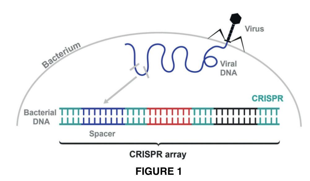
120 Scientific Harrovian 2022
entered the host cell [7]. The long CRISPR RNA is cut by tracRRNA within each repeat into functional units [8]. This binds to the Cas9 protein, a component of CRISPR immunity, to form a surveillance complex [8]. Each small piece of RNA in the cytoplasm contains the genetic information of a previous bacteriophage invader [7]. They provide guidance for an endonuclease (Cas9 protein) that latches onto a matching DNA sequence. It unwinds it and cuts each strand DNA which forms the double helix [7,8]. The broken viral DNA is then cut off and destroyed by other proteins, which stops infection of the bacteria [8].
The mechanism
The CRISPR-Cas9 technology is split into three stages: designing complexes, recognition and interference (cleavage and repair).
1) Designing
Firstly, scientists need to choose a Cas enzyme. The PAM sequence (protospacer-adjacent motif) is important in determining which enzyme will be used as it determines potential target sites for genome editing [14]. There are two main Cas enzymes used in genome editing: Cas9 and Cas12a [14]. To direct the Cas enzyme to the target gene, scientists create a guide RNA (gRNA) which has the complementary sequence to the target gene [10]. The gRNA is either made as a single guide RNA (sgRNA) or a 2-part guide RNA (consisting of both crRNA and tracrRNA). In the Cas12a enzyme, the gRNA is also called crRNA, which is a single molecule of RNA [14].This gRNA is then attached to a Cas9 protein to form a complex ribonucleoprotein (RNP) [10, 14].
2) Recognition
The RNP can then be delivered to the cell by electroporation or lipofection [13]. In the cell, the gRNA with the complementary sequence directs Cas9 to the target gene, which binds precisely to the target gene that needs editing [11,12].
3) Interference (cleavage and repair)
A Cas9 enzyme cuts the genome at the target gene, which forms a specific double-strand break (DSB) at desired locations in the genome [11, 13]. This causes the cell to try to repair the induced break by double-stranded break repair mechanisms (a natural process in bacteria): homology-directed repair (HDR) and non-homologous end joining (NHEJ) [10,15]. In non-homologous end joining (NHEJ), DNA fragments are joined together to repair double strand breaks (DSBs) through an enzymatic process in the absence of exogenous homologous DNA (DNA originating outside an organism that has been introduced into the organism) [15]. This is active in all phases of the cell cycle [15]. However, this process is prone to errors, resulting in unusable genes that are turned off as there is an extra or a missing base, resulting from small random insertion or deletion at cleavage site that led to the generation of frameshift mutation or a premature stop codon [10,15]. HDR requires a separate template DNA to their CRISPR where the cellular protein can perform a different DNA repair process [14]. This template DNA is used to direct the repair and can be used to execute precise gene insertion or replacement by adding a donor DNA template with sequence homology at the predicted DSB site [14]. This is the stage where a short fragment of DNA or the desired gene with a specific function is then inserted to fill the gap and replace the original gene [12].
Applications
Ever since the discovery of CRISPR in 1987, it has been widely applied in humans, bacteria, plants as well as other animals [16]. However, its primary application is in humans - it has recently undergone many clinical trials to test for its effectiveness on eradicating cancer [17]. Cancer is most commonly associated as a terminal illness with no cure; it is caused when cells grow uncontrollably due to mutations and hence, leading to the formation of a tumour [18]. Proto-oncogene is a gene responsible for regulating cell growth during the cell cycle. When proto-oncogene mutates, oncogenes are produced, which are mutated genes that could result in the growth of a tumour [19]. In addition, tumour suppressor genes also play a role in
and
121 Chemistry
Biology
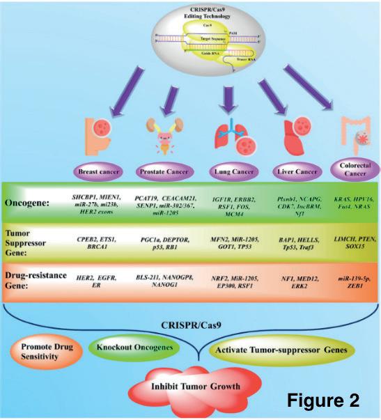
the regulation of cell division. When mutations arise, this could cause the dysfunction of the gene which would lead to deregulation of cell division, resulting in the formation of a tumour [20]. CRISPR gene editing has been undergoing clinical trials and has been shown to be effective in targeting the removal of oncogenes and tumour suppressor genes [21]. Figure 2 depicts the genes that play a role in the development of various types of cancer and the genes that have been successfully targeted during clinical trials.
Limitations of the use of CRISPR gene editing
There are many ethical considerations as well as limitations when using CRISPR gene editing. Concerns revolve around the overall safeness of gene editing [17]. When conducting gene editing on humans, there is a risk of off-targeting effects. Off-targeting effects occur when CRISPR cuts at sites other than the target gene, affecting the function of other genes, changing the proteins that are formed as well as leading to mutations as the base sequence of the gene is altered [22]. To minimise the risk of off-targeting, bioinformatic tools can be used to map out the target gene to see whether there is a high probability of off-targeting occurring [23].
Conclusion
With the CRISPR-Cas9 technology adapted from bacteria’s immune system, scientists are able to change an organism’s DNA, revolutionising the biomedical field. This technology allows scientists to manipulate genes in a precise way by adding, removing or altering genetic information at a specific site in the genome. This can accelerate research into diseases like cancer by removing oncogenes and tumour suppressor genes. Like all techniques, it comes with ethical issues and limitations. Scientists have raised concerns, such as eugenics and the safeness of gene editing, with limitations of off-targeting genes.
References
[1]
Vidyasagar, A. and Lanese, N. (2021). What Is CRISPR? [online] Live Science. Available at: https://www.livescience.com/58790-crispr-explained.html [Accessed 24 Apr. 2023].
[2]
Your Genome (2022). What is CRISPR-Cas9? [online] yourgenome.org. Available at: https://www.yourgenome.org/facts/what-is-crispr-cas9/ [Accessed 24 Apr. 2023].
[3]
CRISPR Therapeutics (2023). CRISPR/Cas9. [online] CRISPR. Available at: https://crisprtx.com/gene-editing/crispr-cas9#:~:text=CRIS-
122 Scientific Harrovian 2022
PR%3A%20Clustered%20Regularly%20Interspaced%20Short [Accessed 24 Apr. 2023].
[4]
medlineplus.gov. (2022). What are genome editing and CRISPR-Cas9?: MedlinePlus Genetics. [online] Available at: https://medlineplus.gov/ genetics/understanding/genomicresearch/genomeediting/#:~:text=The%20CRISPR%2DCas9%20system%20has [Accessed 26 Apr. 2023].
[5]
Arroyo-Olarte, R.D., Bravo Rodríguez, R. and Morales-Ríos, E. (2021). Genome Editing in Bacteria: CRISPR-Cas and Beyond. Microorganisms, [online] 9(4), p.844. doi:https://doi.org/10.3390/microorganisms9040844.
[6]
AddGene (2015). Addgene: CRISPR History and Development for Genome Engineering. [online] Addgene.org. Available at: https://www. addgene.org/crispr/history/ [Accessed 28 Apr. 2023].
[7]
Innovative Genomics Institute – IGI. “CRISPR Immunity Explained: How Cas9 Protects Bacteria from Viruses.” YouTube, 9 Aug. 2021, www. youtube.com/watch?v=Aqw4DihmoQY. Accessed 29 Apr. 2023.
[8]
“How CRISPR Works as a Bacterial Immune System.” Www.youtube.com, 20 Apr. 2021, www.youtube.com/watch?v=aX3fBMXYUTc. Accessed 29 Apr. 2023.
[9]
Wikipedia Contributors. “CRISPR.” Wikipedia, Wikimedia Foundation, 28 Feb. 2019, en.wikipedia.org/wiki/CRISPR. Accessed 29 Apr. 2023.
[10]
TED-Ed. “How CRISPR Lets You Edit DNA - Andrea M. Henle.” YouTube, 24 Jan. 2019, www.youtube.com/watch?v=6tw_JVz_IEc. Accessed 1 May 2023.
[11]
Mayo Clinic. “CRISPR Explained.” YouTube, 24 July 2018, www.youtube.com/watch?v=UKbrwPL3wXE. Accessed 1 May 2023.
[12]
Hasnat Tariq. “Crispr Cas9.” Slideshare, 11 Feb. 2018, www.slideshare.net/hazz12/crispr-cas9-87803136. Accessed 1 May 2023.
[13]
Asit Prasad Dash. “Crispr Cas9.” Slideshare, 16 Sept. 2019, www.slideshare.net/asitpd/crispr-cas9-172435869. Accessed 1 May 2023.
[14]
“CRISPR Genome Editing Workflow .” Integrated DNA Technologies (IDT) , sg.idtdna.com/pages/technology/crispr/crispr-workflow?utm_ source=google&utm_medium=cpc&utm_campaign=00586_1d_03&utm_content=search&gclid=CjwKCAjwxr2iBhBJEiwAdXECw4PUtDCposR_6AZpddKo729As0CE_FkatoVq30tlVa4f_MdwNkrpnRoCzxQQAvD_BwE. Accessed 1 May 2023.
[15]
Asmamaw, Misganaw, and Belay Zawdie. “Mechanism and Applications of CRISPR/Cas-9-Mediated Genome Editing.” Biologics : Targets & Therapy, vol. 15, no. 15, 21 Aug. 2021, pp. 353–361, www.ncbi.nlm.nih.gov/pmc/articles/PMC8388126/, https://doi.org/10.2147/BTT. S326422. Accessed 1 May 2023.
[16] Gostimskaya, I. (2022). CRISPR–Cas9: A History of Its Discovery and Ethical Considerations of Its Use in Genome Editing. Biochemistry (Moscow), [online] 87(8), pp.777–788. doi:https://doi.org/10.1134/s0006297922080090.
[17] Tavakoli, K., Pour-Aboughadareh, A., Kianersi, F., Poczai, P., Etminan, A. and Shooshtari, L. (2021). Applications of CRISPR-Cas9 as an Advanced Genome Editing System in Life Sciences. BioTech, 10(3), p.14. doi:https://doi.org/10.3390/biotech10030014.
[18] Cooper, G.M. (2000). Tumor Suppressor Genes. The Cell: A Molecular Approach. 2nd edition. [online] Available at: https://www.ncbi.nlm. nih.gov/books/NBK9894/#:~:text=Accumulated%20damage%20to%20both%20oncogenes [Accessed 2 May 2023].
[19] National Cancer Institute (2011). https://www.cancer.gov/publications/dictionaries/cancer-terms/def/proto-oncogene. [online] www.cancer. gov. Available at: https://www.cancer.gov/publications/dictionaries/cancer-terms/def/proto-oncogene [Accessed 2 May 2023].
[20] Bell, D.W. (2023). Tumor Suppressor Gene. [online] Genome.gov. Available at: https://www.genome.gov/genetics-glossary/Tumor-Suppressor-Gene#:~:text=%E2%80%8BTumor%20Suppressor%20Gene&text=A%20tumor%20suppressor%20gene%20encodes.
[21] Hazafa, A., Mumtaz, M., Farooq, M.F., Bilal, S., Chaudhry, S.N., Firdous, M., Naeem, H., Ullah, M.O., Yameen, M., Mukhtiar, M.S. and Zafar, F. (2020). CRISPR/Cas9: A powerful genome editing technique for the treatment of cancer cells with present challenges and future directions. Life Sciences, 263, p.118525. doi:https://doi.org/10.1016/j.lfs.2020.118525.
123 Chemistry and Biology
[22] Guo, C., Ma, X., Gao, F. and Guo, Y. (2023). Off-target effects in CRISPR/Cas9 gene editing. Frontiers in Bioengineering and Biotechnology, 11. doi:https://doi.org/10.3389/fbioe.2023.1143157.
[23] Rasul, M.F., Hussen, B.M., Salihi, A., Ismael, B.S., Jalal, P.J., Zanichelli, A., Jamali, E., Baniahmad, A., Ghafouri-Fard, S., Basiri, A. and Taheri, M. (2022). Strategies to overcome the main challenges of the use of CRISPR/Cas9 as a replacement for cancer therapy. Molecular Cancer, 21(1). doi:https://doi.org/10.1186/s12943-021-01487-4.
Figures
Figure 1
Werner, Anina . “What Is CRISPR-Cas9 and How Does It Work? | INTEGRA.” Www.integra-Biosciences.com, 18 Oct. 2022, www.integra-biosciences.com/japan/en/blog/article/what-crispr-cas9-and-how-does-it-work. Accessed 1 May 2023.
Figure 2
Hazafa, A., Mumtaz, M., Farooq, M.F., Bilal, S., Chaudhry, S.N., Firdous, M., Naeem, H., Ullah, M.O., Yameen, M., Mukhtiar, M.S. and Zafar, F. (2020). CRISPR/Cas9: A powerful genome editing technique for the treatment of cancer cells with present challenges and future directions. Life Sciences, 263, p.118525. doi:https://doi.org/10.1016/j.lfs.2020.118525.
124 Scientific Harrovian 2022

The Spanish Flu vs COVID-19
by Branda Mak
125 Chemistry and Biology
Introduction
We are at a stage where COVID-19 is starting to ease. This COVID-19 pandemic has changed lives of people - many in a detrimental way - with rising death tolls of 6.7 million, as well as, global social, political and economic impacts. This has made me wonder what the previous deadly pandemic was like in comparison to COVID-19. This article aims to give a brief history of the Spanish flu and the comparison between the Spanish flu and COVID-19 pandemic.
Overlook of the Spanish Flu
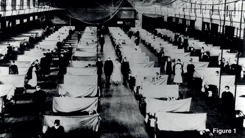
The 1918 influenza pandemic is said to be one of the deadliest pandemics in history. It was caused by an H1N1 Influenza A virus with genes of avian origin, though it is not clear exactly where the virus actually originated [1]. However, most epidemiologists and virologists believe that the earliest documented case is said to be in Kansas, United States [2]. The 1918 Spanish influenza lasted from 1918 to 1920 [1]. The flu spread further with more cases recorded in Germany, France and the United States only after a month from the initial. By the end of the pandemic in April 1920 (two years later), an estimate of 500 million people - nearly a third of the global population at that time - had been infected in four successive waves [1].
The first pandemic wave lasted from February 1918 to June 1918, which was benign and only caused a few deaths. A month of declining infections later, a mutation of the virus caused it to become extremely virulent, leading to millions of deaths globally, lasting from August to December 1918. The third wave was milder, occurring from December 1918 to April 1919 with the fourth wave lasting from December 1919 to April 1920. This data can be seen as a graphic in figure 2.
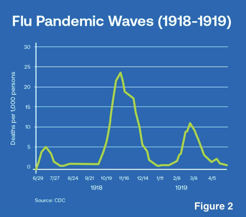
This resulted in an estimated 25 million deaths, with more researchers speculating 40-50 million deaths, affecting young adults from 20 and 40 years of age [1,3]. The outbreak is called the Spanish flu because Spain, a neutral country during WWI, was the first to publicly report cases of the disease. There were already cases in China, France and the United States, but wartime censors suppressed news of the flu to avoid affecting morale [4].
Comparing Patient population
One of the biggest differences between the Spanish Flu and Covid-19 is the patient population. In the Spanish flu, mortality was high in infants under 5 years old, 20-40 years old, and 65 years old or older. The unique feature of this outbreak was the high mortality in healthy people, even within the 20-40 age group [5]. Whereas in Covid-19, ages over 65 was affected the most, especially those with comorbidity [1]. For the 1918 outbreak, the mortality rate for younger people was 8-10% compared with the 2.5% overall mortality rate whereas the mortality rate for the 25-40 age group is a mere 0.2% in contrast to the 2.4% overall mortality rate [6]. Moreover, people aged 25-40 years olds accounted for 40% of deaths from the 1918 influenza, whereas those in the 18-44 year old range accounted for only 3.9% of deaths from Covid-19 [6].
126 Scientific Harrovian 2022
Comparing Virus Type
The Spanish flu was caused by an H1N1 influenza virus, which is an orthomyxovirus. It produces virions that are 80 to 120 nm in diameter, with an RNA genome - sized approx 13.5kb [7]. A virion is the complete virus particle consisting of an RNA core with a protein coat and a capsid. The influenza genome has 8 different regions which are segmented and encodes for 11 different proteins, which are listed down in the table below [7].
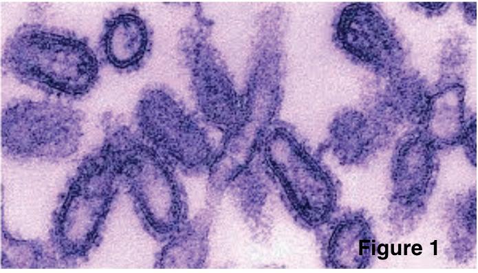
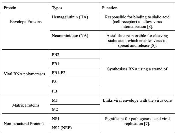
The surface glycoproteins HA and NA are how the H1N1- strain is differentiated from other strains of influenza A (H1N2) depending on the type of HA or NA antigens expressed with metabolic synergy [7].
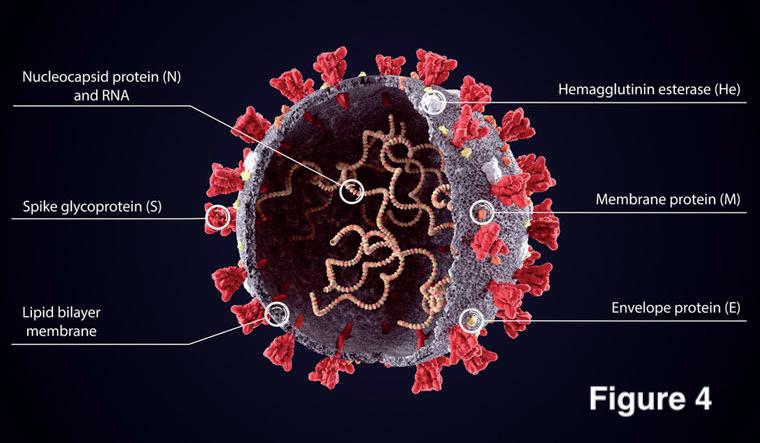
The recent Covid-19 was caused by Severe Acute-Respiratory Syndrome Coronavirus 2 (SARS-CoV-2), which belongs to the β coronavirus subfamily [9,10]. Like other coronaviruses, SARS-CoV-2 has four structural proteins: spike (S), envelope (E), membrane (M), and nucleocapsid (N) proteins [11]. The nucleocapsid protein (N) holds the RNA genome as it forms the capsid outside the genome [11]. Whereas, the spike (S), envelope (E) and membrane (M) proteins creates the viral envelope where the viral genomic RNA is further packed in the envelope [11]. The spike (S) proteins are glycoproteins and is responsible for allowing the virus to attach and fuse with the membrane of a host cell [12]. They are divided into two functional parts (S1 and S2) during their biosynthesis in infected cells [13]. The S1 subunit catalyses the attachment and the S2 subunit anchors the S protein onto the membrane of the host cell [13]. The spike protein is also what gives the crown-like appearance as they are inserted in multiple copies into the membrane of the virion [13].
127 Chemistry and Biology
Comparing Cause of deaths
The two diseases kill via different mechanisms. Deaths from the H1N1 influenza A virus was predominantly due to severe secondary bacterial pneumonia caused by bacterial “pneumopathogens”. These bacterias included pneumococci, streptococci and staphylococci [15]. They colonised the upper respiratory tract common upper respiratory-tract bacteria [14,15]. Pneumonia occurs as bacteria that normally inhabit the nose and throat invades the lung along a pathway created by the virus destroying the cells lining the bronchial tubes and lungs, including cilia [16]. The loss in cells encouraged further loss of cells throughout the entire respiratory tract, including those deep in the lungs [16]. This makes the tract vulnerable to attack by bacteria moving along the newly created pathway from the nose and throat [16].
Those with COVID-19 were killed from an overactive immune response that resulted in multiple organ failure [14]. Researchers found out that severe COVID-19 infection presents with acute respiratory distress syndrome (ARDS) - often also seen with the H1N1 influenza A virus [14]. This causes lung inflammation with the production of thick mucus in bronchioles, clogging the airflow which damages both lungs [17]. Due to the severe lung infections, patients experience a widespread formation of blood clots in the body [17]. This makes it challenging for the patient to breathe on their own, so they require invasive mechanical ventilation [17]. This means that a tube needs to inserted in the airway connected to a ventilator to help patients breathe. Among them, a large number of patients die. This severity results from overactive neutrophils) [18]. Neutrophils are white blood cells that detect bacteria and respond by expelling their DNA laced with NET to attack the bacteria[18]. NET stands for neutrophil extracellular trap and is a toxic enzyme which digests unwanted pathogens [18]. High concentration of NETs positively correlates with sepsis severity and organ dysfunction, as well as contributing to immunothrombosis (a network between the coagulation and innate immune system) during inflammatory response [19]. Moreover, innate immune cells - involved in the body’s first immune response to a harmful foreign substance - induce endothelial damage, acute lung injury, disruption of lung structure, multi-organ damage which may all lead to deaths [19].
Comparing Vaccinations
For both cases, treatments and vaccines were found. Though it took a shorter time to develop COVID-19 vaccine as more knowledge was gained over the last century [20]. The initial treatment for the Spanish flu was the non-pharmaceutical interventions, such as isolation, quarantine, disinfectant and limitations of public gatherings - similar to COVID-19 [21]. Many vaccines were produced to try to prevent influenza symptoms. They were all developed based on Pfeiffer’s bacillus - the probable cause of influenza. William H. Park succeeded in developing a vaccine that combated diphtheria with anti-toxins [22]. After this vaccine, faculty from the Medical School of the University of Pittsburgh produced a vaccine by modifying Park’s techniques [22]. The vaccine was then tested for toxicity and distributed to the Red Cross for patients [22].
For the first two years of the COVID-19 pandemic, there were no specific and effective treatments or cures available to patients [23]. The first COVID-19 vaccine to be approved by the US Food and Drug Administration was the Pfizer-BioNTech vaccine for individuals 16 years old or older in August 2021 [24]. Since then, more and more COVID-19 vaccines were introduced - including Moderna, Novavax and Johnson & Johnson’s Jannsen (J&J/Janssen) [25]. Data from February 2021 showed that Pfizer-BioNTech was the most popular vaccine with 61 countries using them [28].
The Pfizer-BioNTech and Moderna are mRNA vaccines. The mRNA vaccine causes our bodies to produce antibodies as part of the immune response. This will fit onto surface proteins of the coronavirus to prevent it from entering the host cell. The mRNA in vaccines are wrapped in a protective layer of fat which helps it get taken up by dendritic cells (specialised cell in the immune system) [27]. Once inside the cells, the mRNA attaches to a ribosome, which produces pieces of viral surface protein(antigen) - usually found on
128 Scientific Harrovian 2022
the virus’ outer membrane [26]. This are then displayed on the surface of dendritic cells [27]. The dendritic cell travels to nearby lymph node where it presents the antigen proteins to other cells of the immune system, including helper T cells. The helper T cells teach B cells to make antibodies that fit onto the surface protein of coronavirus [27]. Other cells, such as cytotoxic T cells, are stimulated by the protein piece and can kill virus infected cells [27]. These antibodies stay within our body. When the body is exposed to the coronavirus, the immune system is able to recognise, neutralise and destroy it.
Conclusion
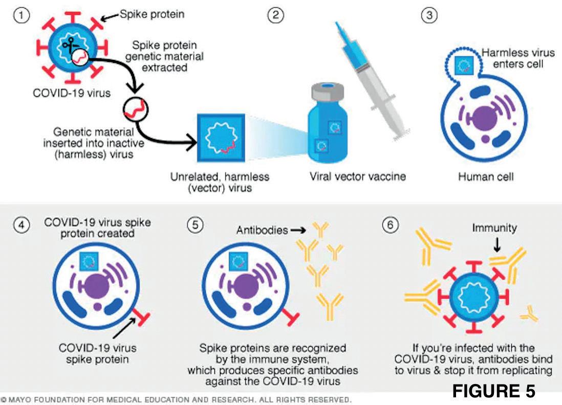
The two pandemics differed in causes of disease, mechanism of death and the risk population. H1N1 influenza A virus caused the Spanish flu, whereby most victims died from secondary bacterial pneumonia. Whereas, SARS-CoV-2 (a form of betacoronavirus) was the cause of COVID-19 and victims mostly died due to the overactive immune response resulting in organ failure. The 1918 influenza affected less than half of all countries, where the most vulnerable groups were healthy adults between 25 - 40 years old. Meanwhile, COVID-19 affected nearly all countries and the most vulnerable groups are the elderly, aged 65 and above with comorbidities. Acute respiratory distress syndrome (ARDS) can develop in both cases, however, in influenza ARDS resulted in 100% fatality rate compared to the 53.4% mortality rate as a complication from COVID-19. Moreover, both pandemics had a delay in treatment and vaccine. The most popular vaccine used worldwide is the Pfizer-BioNTech vaccine which uses mRNA to provide immunity from the coronavirus.
References
[1]
Liang, Shu Ting, et al. “COVID-19: A Comparison to the 1918 Influenza and How We Can Defeat It.” Postgraduate Medical Journal, vol. 97, no. 1147, 9 Feb. 2021, pmj.bmj.com/content/early/2021/02/08/postgradmedj-2020-139070, https://doi.org/10.1136/postgradmedj-2020-139070. Accessed 25 Apr. 2023.
[2]
Wikipedia Contributors. “Spanish Flu.” Wikipedia, Wikimedia Foundation, 25 Oct. 2019, en.wikipedia.org/wiki/Spanish_flu. Accessed 25 Apr. 2023.
[3]
Erkoreka, Anton. “| 190-194 | OPEN ACCESS.” Journal of Molecular and Genetic Medicine, vol. 3, no. 2, 2009, pp. 190–194, citeseerx.ist.psu. edu/document?repid=rep1&type=pdf&doi=aa151f11ecdcf9c58087e1f89f8f5b7240a82888. Accessed 25 Apr. 2023.
[4]
Brown, Matthew. “Fact Check: Why Is the 1918 Influenza Virus Called “Spanish Flu”?” USA TODAY, 23 Mar. 2020, www.usatoday.com/story/ news/factcheck/2020/03/23/fact-check-how-did-1918-pandemic-get-name-spanish-flu/2895617001/. Accessed 24 Apr. 2023.
[5]
Centers for Disease Control and Prevention. “1918 Pandemic (H1N1 Virus) .” Cdc.gov, Centers for Disease Control and Prevention, 20 Mar. 2019, www.cdc.gov/flu/pandemic-resources/1918-pandemic-h1n1.html . Accessed 27 Apr. 2023.
[6]
Worldometer. “Coronavirus Age, Sex, Demographics (COVID-19) - Worldometer.” Www.worldometers.info, 13 May 2020, www.worldometers. info/coronavirus/coronavirus-age-sex-demographics/. Accessed 27 Apr. 2023.
[7]
Jilani, Talha N., et al. “H1N1 Influenza (Swine Flu).” PubMed, StatPearls Publishing, 2020, www.ncbi.nlm.nih.gov/books/NBK513241/#:~:text=The%20H1N1%20influenza%20virus%20is. Accessed 27 Apr. 2023.
[8]
Benton, Donald J., et al. “Role of Neuraminidase in Influenza A(H7N9) Virus Receptor Binding.” Journal of Virology, vol. 91, no. 11, 1 June 2017, jvi.asm.org/content/91/11/e02293-16, https://doi.org/10.1128/JVI.02293-16. Accessed 27 Apr. 2023.
[9]
WHO. “Naming the Coronavirus Disease (COVID-19) and the Virus That Causes It.” World Health Organization, 2020, www.who.int/emer129 Chemistry and Biology
gencies/diseases/novel-coronavirus-2019/technical-guidance/naming-the-coronavirus-disease-(covid-2019)-and-the-virus-that-causes-it. Accessed 27 Apr. 2023.
[10]
Ahmadpour , Doryaneh , and Pedram Ahmadpoor. “How the COVID-19 Overcomes the Battle? An Approach to Virus Structure .” Www.ijkd.org, Apr. 2020, www.researchgate.net/profile/Pedram-Ahmadpoor/publication/341131911_How_the_COVID-19_Overcomes_the_Battle_An_Approach_to_Virus_Structure/links/5f281f63299bf134049cf33c/How-the-COVID-19-Overcomes-the-Battle-An-Approach-to-Virus-Structure.pdf. Accessed 27 Apr. 2020.
[11]
Wang, Mei-Yue, et al. “SARS-CoV-2: Structure, Biology, and Structure-Based Therapeutics Development.” Frontiers in Cellular and Infection Microbiology, vol. 10, no. 587269, 25 Nov. 2020, pubmed.ncbi.nlm.nih.gov/33324574/, https://doi.org/10.3389/fcimb.2020.587269. Accessed 27 Apr. 2023.
[12]
Wu, Canrong, et al. “Analysis of Therapeutic Targets for SARS-CoV-2 and Discovery of Potential Drugs by Computational Methods.” Acta Pharmaceutica Sinica B, vol. 10, no. 5, Feb. 2020, https://doi.org/10.1016/j.apsb.2020.02.008. Accessed 28 Apr. 2023
[13]
Jackson, Cody B., et al. “Mechanisms of SARS-CoV-2 Entry into Cells.” Nature Reviews Molecular Cell Biology, vol. 23, no. 1, 5 Oct. 2021, pp. 1–18, https://doi.org/10.1038/s41580-021-00418-x. Accessed 28 Apr. 2023.
[14]
---. “COVID-19: A Comparison to the 1918 Influenza and How We Can Defeat It.” Postgraduate Medical Journal, vol. 97, no. 1147, 9 Feb. 2021, pmj.bmj.com/content/early/2021/02/08/postgradmedj-2020-139070, https://doi.org/10.1136/postgradmedj-2020-139070. Accessed 28 Apr. 2023.
[15]
Morens, David M., et al. “Predominant Role of Bacterial Pneumonia as a Cause of Death in Pandemic Influenza: Implications for Pandemic Influenza Preparedness.” The Journal of Infectious Diseases, vol. 198, no. 7, Oct. 2008, pp. 962–970, https://doi.org/10.1086/591708. Accessed 28 Apr. 2023.
[16]
“Bacterial Pneumonia Caused Most Deaths in 1918 Influenza Pandemic.” National Institutes of Health (NIH), 23 Sept. 2015, www.nih.gov/ news-events/news-releases/bacterial-pneumonia-caused-most-deaths-1918-influenza-pandemic. Accessed 28 Apr. 2023.
[17]
Thomas, Dr. Liji. “Are Overactive Immune Cells Causing COVID-19-Related Deaths?” News-Medical.net, 16 Apr. 2020, www.news-medical.net/ news/20200416/Are-overactive-immune-cells-causing-COVID-19-related-deaths.aspx. Accessed 28 Apr. 2023.
[18]
Laboratory, Cold Spring Harbor. “Are Overactive Immune Cells the Cause of COVID-19 Deaths?” SciTechDaily, 16 Apr. 2020, scitechdaily.com/ are-overactive-immune-cells-the-cause-of-covid-19-deaths/. Accessed 29 Apr. 2023.
[19]
Li Yin Tan, et al. “Hyperinflammatory Immune Response and COVID-19: A Double Edged Sword.” Frontiers - Frontiers in Immunology, 30 Sept. 2021, www.frontiersin.org/articles/10.3389/fimmu.2021.742941/full#:~:text=It%20involves%20activation%20of%20multiple,health%20 effects%20of%20COVID%2D19. Accessed 29 Apr. 2023.
[20]
Hanan, Mishayl. “COVID-19 and the Spanish Flu - Drawing Comparisons.” BioSpace, 19 Oct. 2021, www.biospace.com/article/covid-19-andthe-spanish-flu-drawing-comparisons-/. Accessed 29 Apr. 2023. [21]
---. “History of 1918 Flu Pandemic.” Centers for Disease Control and Prevention, CDC, 21 Mar. 2018, www.cdc.gov/flu/pandemic-resources/1918-commemoration/1918-pandemic-history.htm. Accessed 29 Apr. 2023. [22]
Eyler, John M. “The State of Science, Microbiology, and Vaccines circa 1918.” Public Health Reports, vol. 125, no. 3_suppl, Apr. 2010, pp. 27–36, www.ncbi.nlm.nih.gov/pmc/articles/PMC2862332/, https://doi.org/10.1177/00333549101250s306. Accessed 1 May 2023. [23]
Siemieniuk, Reed Ac, et al. “Drug Treatments for Covid-19: Living Systematic Review and Network Meta-Analysis.” BMJ (Clinical Research Ed.), vol. 370, 30 July 2020, p. m2980, pubmed.ncbi.nlm.nih.gov/32732190/, https://doi.org/10.1136/bmj.m2980. Accessed 1 May 2023.
[24]
FDA. “FDA Approves First COVID-19 Vaccine.” FDA, 23 Aug. 2021, www.fda.gov/news-events/press-announcements/fda-approves-first-covid19-vaccine. Accessed 1 May 2023.
[25]
CDC. “COVID-19 and Your Health.” Centers for Disease Control and Prevention, 11 Feb. 2020, www.cdc.gov/coronavirus/2019-ncov/vaccines/ stay-up-to-date.html. Accessed 1 May 2023.
[26]
MedlinePlus. “What Are MRNA Vaccines and How Do They Work?: MedlinePlus Genetics.” Medlineplus.gov, 21 Nov. 2022, medlineplus.gov/ genetics/understanding/therapy/mrnavaccines/. Accessed 1 May 2023.
[27]
“How COVID-19 MRNA Vaccines Work.” Www.youtube.com, 29 July 2021, www.youtube.com/watch?v=8nD6Q9X0SFw. Accessed 1 May 2023. [28]
McCarthy, Niall. “Infographic: Which Covid-19 Vaccines Are Most Widely Used?” Statista Infographics, 16 Feb. 2021, www.statista.com/ chart/24191/number-of-countries-using-selected-covid-19-vaccines/. Accessed 1 May 2023.
Figures
Figure 1
History.com Editors. “Spanish Flu.” History, A&E Television Networks, 12 Oct. 2010, www.history.com/topics/world-war-i/1918-flu-pandemic.
130 Scientific Harrovian 2022
Accessed 29 Apr. 2023.
www.theweek.in/news/health/2020/06/30/xrays-size-up-coronavirus-structure-at-room-temperature.html. Accessed 28 Apr. 2023.
Figure 2
https://www.google.com.hk/url?sa=i&url=https%3A%2F%2Ftheconversation.com%2Fcompare-the-flu-pandemic-of-1918-and-covid-19with-caution-the-past-is-not-a-prediction-138895&psig=AOvVaw08f9dILRKuYAvrNY5Znr3W&ust=1686643497299000&source=images&cd=vfe&ved=0CBEQjRxqFwoTCNDwk-aivf8CFQAAAAAdAAAAABAE
Figure 3
History.com Editors. “Spanish Flu.” History, A&E Television Networks, 12 Oct. 2010, www.history.com/topics/world-war-i/1918-flu-pandemic. Accessed 20 June 2023.
Figure 4
“X-Rays Size up Coronavirus Protein Structure at Room Temperature.” The Week, 30 June 2020, Figure 5
Mayo Clinic. “How Do Different Types of COVID-19 Vaccines Work?” Mayo Clinic, 5 Nov. 2021, www.mayoclinic.org/diseases-conditions/coronavirus/in-depth/different-types-of-covid-19-vaccines/art-20506465. Accessed 20 June 2023.
Citations
https://pmj.bmj.com/content/97/1147/273
https://en.wikipedia.org/wiki/Spanish_flu
https://citeseerx.ist.psu.edu/document?repid=rep1&type=pdf&doi=aa151f11ecdcf9c58087e1f89f8f5b7240a82888
https://www.usatoday.com/story/news/factcheck/2020/03/23/fact-check-how-did-1918-pandemic-get-name-spanish-flu/2895617001/ https://www.cdc.gov/flu/pandemic-resources/1918-pandemic-h1n1.html
https://www.worldometers.info/coronavirus/coronavirus-age-sex-demographics/
https://www.ncbi.nlm.nih.gov/books/NBK513241/#:~:text=The%20H1N1%20influenza%20virus%20is,HA)%20and%20neuraminidase%20(NA)
https://journals.asm.org/doi/10.1128/JVI.02293-16
https://www.who.int/emergencies/diseases/novel-coronavirus-2019/technical-guidance/naming-the-coronavirus-disease-(covid-2019)-and-the-virus-that-causes-it
https://www.researchgate.net/profile/Pedram-Ahmadpoor/publication/341131911_How_the_COVID-19_Overcomes_the_Battle_An_Approach_to_Virus_Structure/links/5f281f63299bf134049cf33c/How-the-COVID-19-Overcomes-the-Battle-An-Approach-to-Virus-Structure.pdf
https://www.ncbi.nlm.nih.gov/pmc/articles/PMC7723891/
https://www.ncbi.nlm.nih.gov/pmc/articles/PMC7102550/
https://www.ncbi.nlm.nih.gov/pmc/articles/PMC8491763/
https://www.ncbi.nlm.nih.gov/pmc/articles/PMC8108277/ https://academic.oup.com/jid/article/198/7/962/2192118
https://www.nih.gov/news-events/news-releases/bacterial-pneumonia-caused-most-deaths-1918-influenza-pandemic
https://www.news-medical.net/news/20200416/Are-overactive-immune-cells-causing-COVID-19-related-deaths.aspx https://scitechdaily.com/are-overactive-immune-cells-the-cause-of-covid-19-deaths/
https://www.frontiersin.org/articles/10.3389/fimmu.2021.742941/full#:~:text=It%20involves%20activation%20of%20multiple,health%20effects%20of%20 COVID%2D19.
https://www.biospace.com/article/covid-19-and-the-spanish-flu-drawing-comparisons-/ https://www.cdc.gov/flu/pandemic-resources/1918-commemoration/1918-pandemic-history.htm
https://www.ncbi.nlm.nih.gov/pmc/articles/PMC2862332/#:~:text=Many%20vaccines%20were%20developed%20and,results%20of%20these%20vaccine%20 trials.
https://www.ncbi.nlm.nih.gov/pmc/articles/PMC7390912/
https://www.fda.gov/news-events/press-announcements/fda-approves-first-covid-19-vaccine
https://www.cdc.gov/coronavirus/2019-ncov/vaccines/stay-up-to-date.html
https://medlineplus.gov/genetics/understanding/therapy/mrnavaccines/
https://www.youtube.com/watch?v=8nD6Q9X0SFw
https://www.statista.com/chart/24191/number-of-countries-using-selected-covid-19-vaccines/
https://citeseerx.ist.psu.edu/document?repid=rep1&type=pdf&doi=aa151f11ecdcf9c58087e1f89f8f5b7240a82888
https://www.usatoday.com/story/news/factcheck/2020/03/23/fact-check-how-did-1918-pandemic-get-name-spanish-flu/2895617001/ https://www.cdc.gov/flu/pandemic-resources/1918-pandemic-h1n1.html
https://www.worldometers.info/coronavirus/coronavirus-age-sex-demographics/
https://www.ncbi.nlm.nih.gov/books/NBK513241/#:~:text=The%20H1N1%20influenza%20virus%20is,HA)%20and%20neuraminidase%20(NA) https://journals.asm.org/doi/10.1128/JVI.02293-16
https://www.who.int/emergencies/diseases/novel-coronavirus-2019/technical-guidance/naming-the-coronavirus-disease-(covid-2019)-and-the-virus-that-causes-it https://www.researchgate.net/profile/Pedram-Ahmadpoor/publication/341131911_How_the_COVID-19_Overcomes_the_Battle_An_Approach_to_Virus_Structure/links/5f281f63299bf134049cf33c/How-the-COVID-19-Overcomes-the-Battle-An-Approach-to-Virus-Structure.pdf https://www.ncbi.nlm.nih.gov/pmc/articles/PMC7723891/
https://www.ncbi.nlm.nih.gov/pmc/articles/PMC7102550/
https://www.ncbi.nlm.nih.gov/pmc/articles/PMC8491763/ https://www.ncbi.nlm.nih.gov/pmc/articles/PMC8108277/
https://academic.oup.com/jid/article/198/7/962/2192118
https://www.nih.gov/news-events/news-releases/bacterial-pneumonia-caused-most-deaths-1918-influenza-pandemic
https://www.news-medical.net/news/20200416/Are-overactive-immune-cells-causing-COVID-19-related-deaths.aspx https://scitechdaily.com/are-overactive-immune-cells-the-cause-of-covid-19-deaths/
https://www.frontiersin.org/articles/10.3389/fimmu.2021.742941/full#:~:text=It%20involves%20activation%20of%20multiple,health%20effects%20of%20 COVID%2D19.
https://www.biospace.com/article/covid-19-and-the-spanish-flu-drawing-comparisons-/ https://www.cdc.gov/flu/pandemic-resources/1918-commemoration/1918-pandemic-history.htm
https://www.ncbi.nlm.nih.gov/pmc/articles/PMC2862332/#:~:text=Many%20vaccines%20were%20developed%20and,results%20of%20these%20vaccine%20 trials.
https://www.ncbi.nlm.nih.gov/pmc/articles/PMC7390912/
https://www.fda.gov/news-events/press-announcements/fda-approves-first-covid-19-vaccine
https://www.cdc.gov/coronavirus/2019-ncov/vaccines/stay-up-to-date.html
https://medlineplus.gov/genetics/understanding/therapy/mrnavaccines/
https://www.youtube.com/watch?v=8nD6Q9X0SFw
https://www.statista.com/chart/24191/number-of-countries-using-selected-covid-19-vaccines/
131 Chemistry and Biology

Social Prescribing
by Katy Shiu and Charlize Mui
132 Scientific Harrovian 2022
Introduction
An idea that grew from an impossible, seemingly insane dream has now developed into a solution for our health - a type of solution that may even be considered the most effective against all types of diseases, including those we are unaware of.
This is accomplished through social prescribing. But first before we delve deeper, let us define what social prescribing is. What do most people think the word “health” means? A staggering 93% of the general public believe health is a subject related to biomedicine, such as medicine, pills and vaccines, whereas only 7% of people think of health as a concept in terms of lifestyle choices and prevention, such as exercise, nutrition, and sleep. Such Perception of health is arguably far from the truth. Health does not begin in hospitals.It begins in our neighbourhoods and in our homes.
What is social prescribing?
Recent studies show that 80% of a person’s health is influenced by non-medical factors such as income, access to services, food and clean water, race, mental well-being and social health, whilst only 20% of a person’s health is determined by the result of clinical care. From this study, it can be seen that health starts within our communities.
In contrast to traditional medicine, which prescribes drugs to patients, social prescribing broadens the options available to GPs and other community-based practitioners by providing individualised care for people’s physical and mental health through social interventions. Activities such as dance classes, meditation, art therapy, gardening groups, or community projects may be included, as well as services such as debt counselling, housing advice, or employment assistance. Social prescribing is an approach that aims to improve health and wellbeing while reducing inequalities and optimising the use of healthcare services. It Improves individual health and also lowers an individual’s overall healthcare costs.
Furthermore, as a result of an ageing population in today’s society, there is an increase in complex health and social needs. Additionally, an increase in demand for services is leading to the rising popularity of social prescribing. Many national organisations and individuals from policy, practise, and academia,such as NHS England, the RCGP, The Mayor of London, and the National Institute for Health Research, are rightly advocating for this new option.
Rather than simply treating the symptoms of a specific medical condition and administering the drug required to deal with the diagnosis, the idea is to address the fundamental social, economic, and environmental factors that can contribute to poor health.
Social prescribing was developed in the United Kingdom, with plans and systems dating back decades. For example, Bromley by Bow Health Partnership general practitioners launched a social prescribing scheme in which they referred patients to in-house expert non-clinical services. Similar models of service provision existed in other countries, but many of them were not grouped under the umbrella term of social prescribing.
Components of social prescribing
Since social prescribing programmes often consist of numerous components, namely assessment, referral, coordination, and follow up, it can take some time for benefits to be noticed. This section will concentrate on the key components included in social prescribing programmes.
The assessment is the initial step in the social prescribing process. Patients are examined to identify their social determinants of health, and appropriate non-medical services and resources are chosen to best address the patient’s needs. Many instruments and methods, including surveys, interviews and questionnaires, may be used in this assessment. Healthcare providers can collaborate with community-based organ133 Chemistry and Biology
isations to locate the services and resources available.
Referral is the second component of social prescribing. After the patient has been thoroughly assessed, they are referred to community-based resources and programmes that assist them in addressing their social determinants of health. such as Food banks, housing assistance programmes, job training programmes. Community-based activities such as exercise classes, art programmes, and group therapy are examples. These can also be good options for patients who would like an opportunity to socialise. The goal of referral is to inform patients about available resources or to make direct referrals to community-based organisations.
Coordination is the program’s third component. Healthcare providers collaborate with community-based organisations to provide coordinated care and assistance to patients in need. This may entail forming partnerships between healthcare providers and community-based organisations, or forming care teams composed of both healthcare providers and staff from community-based organisations. Coordination in social prescribing programmes can also refer to the sharing of information among healthcare providers to ensure that patients receive comprehensive care.
Lastly, follow-up is the fourth component of social prescribing. Regular check-ins with health care providers and organisation staff allow them to monitor patient progress and outcomes, ensuring that the patient is receiving the support they require, and making adjustments to their care plan if needed. Follow-up is regarded as one of the most important components of social prescribing because it ensures that the patient is receiving the appropriate long-term support to address their social determinants of health and, ultimately, improve their health outcomes.
Benefits of social prescribing
Social prescribing has a range of benefits for patients, healthcare providers, and society as a collective. Benefits can range from improving social cohesion to building community resilience through promoting community-based activities and services. This is done by connecting patients to non-medical resources in their local communities, such as community groups, charities and social enterprises. By doing so, it helps to build a stronger, more tightly-knit community with a greater sense of solidarity among its members, as well as a network of support.
Another advantage is the reduction of healthcare costs. External factors are the roots of health problems. Patients who participate in social prescribing programmes are less likely to require medical treatments and hospitalisation in the future. It saves costs for both the individual and the healthcare system.
Health inequality is a major issue that can be addressed within the healthcare system through social prescribing. By connecting patients to resources that they might not otherwise have access to, social prescribing reduces disparities in health outcomes between different socioeconomic groups. .
Apart from having an impact on a societal scale, social prescribing can have a variety of positive influences on an individual level. It enables people to have increased control over their own health. Social prescribing has distinct links to self-determination, including autonomy, and the need to have close and affectionate relationships, which fosters a sense of belonging, competence, and beneficence. This proves to be a beneficial method of medical prescription and enables the population to overcome significant socioeconomic inequalities and weak social safety nets, improving an individual’s health drastically.
134 Scientific Harrovian 2022

Social prescribing provides a more individualised approach to care unlike traditional biomedical approaches. This is accomplished by tailoring activities and services to the patient’s specific needs and interests, which is an alternative to the healthcare industry’s one-size-fits-all approach. As a result, patients are happier with their healthcare services.
Finally, social prescribing has a significant impact on reducing social isolation. By connecting patients to community-based activities and services, such programmes can help to reduce loneliness. For example, a patient who is experiencing social isolation as a result of financial difficulties may be referred to a debt counselling service, improving the patient’s mental health and social support networks.
Overall, social prescribing can have a variety of societal and personal benefits. It provides a more holistic and personalised approach to healthcare that can improve patient outcomes while also lowering healthcare costs by addressing the underlying social, economic, and environmental factors that can contribute to poor mental and physical health in the first place.
How has social prescribing changed society so far?
The effect of social prescription gives a clear indication that prevention is potentially of a greater significance to health than clinical care itself. This is because as the population increases, so will the number of people developing chronic diseases. In spite of this, the number of doctors will stay the same, which puts further pressure on healthcare services and increases the risk of global health decline.Therefore, social prescribing changes the society’s perception of the healthcare system from a biomedical model to a bio-psycho-social model, alleviating the pressure on the system. For example, the encouragement of prevention within the healthcare system has been seen to reduce emergency admissions by 14% in Shropshire and Frome alone. It has reduced appointments in the clinic by 3.4 million, saving 300 million pounds which can be reinvested in the healthcare system globally. This allows for the addressing of the people’s needs in a holistic manner.
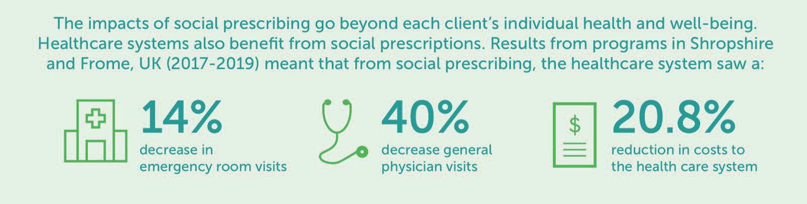
135 Chemistry and Biology
This is evident in a number of successful social prescribing programmes like the Bromley-by-Bow centre in London, a healthcare facility focused on supporting people through a wide range of integrated services tailored to their specific needs. This centre includes a social prescribing service. It provides community-based activities like arts, gardening, and creative activities, as well as services like healthy lifestyle advice, physical activity, employment, volunteering, and counselling. The centre aims to improve these areas in order for patients to make positive life changes. Furthermore, this social prescribing programme collaborates closely with local healthcare providers, assisting in the referral of patients to appropriate activities and services. A study of the programme found that it was closely associated with the reduction of healthcare cost and improved patient outcome.
Another example to mention is the Green Dreams Project in Sheffield, UK. This project introduces the support of tailored coordinated care by combining health needs and social needs through the creation of meaningful group activities. In this project, they employ project managers which link patients to individual GP practices. Using this method, the GP is able to spend time with each patient in order to define the barriers to enhanced quality of life. This programme is designed to be specific to each patient to address these difficulties. Not to mention, this project is also awarded as the runner up for the College of Medicine’s 2013 Innovations Award.
How does social prescribing fit in with wider health and care policy?
Social prescribing and other similar approaches have been used in the NHS for many years. Some schemes dating back to the 1990s or even earlier (e.g. the Bromley-by-Bow Centre was established in 1984). However, for a long time, social prescribing was practised in isolated groups, largely unnoticed by NHS bodies.
In recent years, there has been a significant shift: NHS bodies have embraced social prescribing and committed resources to implementing it across England. The NHS five-year forward view (2014) opened the door to social prescribing by emphasising prevention, the role of the voluntary and community sector, and citing examples of successful social prescribing schemes. Following that, the General Practice Forward View (2016) highlighted the importance that social prescribing holds in providing people with community-based support alongside GP services.
By incorporating social prescribing into its comprehensive model of personalised care, the NHS long-term plan (2019) marked a step change in ambition. The model, which consists of six programmes, including individualised care planning and personal health budgets, aims to empower people, particularly those with more complex needs, to take greater control of their health and care.
Primary care networks (PCNs), groups of GP surgeries serving populations of around 30,000 to 50,000 patients, serve as the channel for this resource and will often host the link-worker service, where a trained social prescribing worker works to identify what is affecting someone’s health and wellbeing. . A new fiveyear contract framework for general practices went into effect in 2019, allowing any PCN with a population of 30,000 or more to be reimbursed for the costs of employing a link worker (one full-time equivalent and more for PCNs with populations of more than 100,000). NHS bodies reported that more than 1,200 link workers were in place by autumn in 2020.
NHS organisations are also working to expand the infrastructure that facilitates social prescribing in addition to funding link workers. Following this, the National Academy of Social Prescribing received £5 million from the Department of Health and Social Care in 2019 to launch. With assistance from a number of partner organisations, including NHS England, NHS Improvement, and Sport England, the academy was formally established as an independent charity in 2020. The academy intends to concentrate on increasing awareness of social prescribing, developing the evidence base, and disseminating promising practices or methods. In addition, it looks for funding alliances and supports nonprofit organisations engaged in social
136 Scientific Harrovian 2022
prescribing.
In recent years, interest in the possibilities of non-clinical interventions has grown amongst other governmental departments. The implementation of social prescribing was supported by the government’s strategy to combat loneliness in 2018. In 2020, the Department for Environment, Food, and Rural Affairs announced funding for a two-year trial of “green social prescribing” initiatives, which are meant to encourage people to interact with the natural world.
The effect of social prescribing globally
The BMC Health Services Research focused on a social prescribing programme in Northern England that uses “Wellbeing Coordinators” to support individuals by offering them information on neighbourhood organisations and services. The purpose of the study was to comprehend both the service’s results and the delivery procedures. At the initial and post-stage, 342 individuals submitted comprehensive and detailed data on their wellbeing and had 26 semi-structured qualitative interviews conducted. From this data, it was shown that participants’ perceived levels of health, social connectivity, and wellbeing had improved, and their levels of anxiety had decreased. However, the data on future access to primary care being restricted was inconclusive. It suggested that men may gain more from social prescribing than women and that a robust and active voluntary and community sector was one of the major factors that boosted the likelihood of success on the social prescribing programme. Another was the maintained and adaptable relationship between the service user and the wellbeing coordinator.
From this research, it was concluded that there was potential for social prescribing to address the social and health requirements of people and communities. This study has found that when service consumers interact with the service, a variety of good effects occur. It is important to think about social prescribing as a means to complement basic care and address unmet needs. This suggests that so long as the patient is willing to interact and be involved with the prescribed initiative’s process, social prescribing programmes can be applicable worldwide.
Social prescribing has even spread its influence as far as the global market, resulting in a day specifically to celebrate and spread awareness about this form of prescription. This day was held on the 9th of March this year and included an event held by the Canadian Institute for Social prescribing. They invited 11 different specialist speakers to give an informative talk through an online webinar. Although social prescribing day isn’t yet on our calendars, it is held annually in March and allows the promotion of this form of medical prescription.
Additionally, the World Health Organisation published a “Toolkit on how to implement social prescribing” which documents 7 steps on how to implement social prescribing, referencing a variety of research and sources. These steps include conducting a situation analysis, assembling a core implementation team, developing an implementation work plan, mapping out community resources, getting everyone on board, link worker training, and monitoring and evaluation. This document addresses each step in detail, i.e. what to consider for situation analysis like the population, feasibility, network and stakeholders. It includes case studies and real life examples for each section.
Challenges and Limitations
Concerning the difficulties of social prescribing, issues frequently revolve around how this newer method of treatment is novel, different, and limited in many ways.
For starters, there is insufficient research and data on this method. Even though the concept can be traced back decades, social prescribing has only recently been recognised in previous years, implying that there is still a lack of rigorous research to support its outcomes. Healthcare providers struggle to determine whether social prescribing intervention is effective for their patients in the absence of conclusive and substantial
137 Chemistry and Biology
evidence.
Furthermore, because social prescribing programmes provide a wide range of treatments, this can arguably be limiting in some ways. As with a variety of interventions (such as exercise classes, art programmes, and community projects), collecting sufficient evidence to support the effectiveness of these interventions requires a long period of research and observation. As a result, tailoring these treatments to specific needs and preferences is difficult. While the concept of social prescribing is gaining popularity, determining which healthcare intervention is most efficient for both the patient’s health and well-being, and how they can be tailored to individual patient needs remains a challenge.
Moreover, as previously stated, this concept has only recently begun to be implemented, programmes that are not yet widely available, land access can be limited for certain populations, such as those living in rural or remote areas. The availability of social prescribing programmes also presents challenges for patients to procure the assistance they require, limiting the reach of such schemes.
Fourthly, financial constraints contribute significantly to the limitations and challenges of social prescribing programmes. Because programmes primarily rely on external funding sources, such as grants or donations, to support their operations, they frequently face a variety of challenges. There is a challenge of limited resources because social prescribing programmes must have the resources to provide services to all patients who need them, particularly in areas with high levels of need.
In relation to this, the reason for limited resources may also be due to funding inequities. Funding for social prescribing programmes may be distributed differently or unfairly if some areas or populations receive more funding than others. As a result, there may be insecure funding. Funding for such programmes can be unpredictable and inconsistent, making it difficult to plan and sustain long-term initiatives.
Conclusion
In conclusion, social prescribing may be regarded as an effective strategy for resolving healthcare challenges in modern society. Social prescribing is a potentially beneficial method of healthcare that has grown in popularity and acceptance in recent years. Projects such as Bromley-By-Bow and Green Dreams Project have the potential to strengthen community bonds, improve patient outcomes, and reduce healthcare costs. However, there are undoubtedly many challenges and constraints associated with social prescribing, such as a lack of sufficient data, financial constraints, and a lack of integration with current conventional healthcare services.
Despite these challenges, social prescribing has been promising in society, and it has shown to improve patient health outcomes and reduce health disparities. As social prescribing continues to grow, healthcare systems will continue to recognise the importance of addressing social determinants of health, leading to a greater likelihood for social prescribing programmes to become more widely implemented and rigorously evaluated.
To ensure the success of social prescribing programmes, it is crucial to implement social prescribing on a national scale while paying close attention to learning. For instance, more the effects of various link working models and how link workers can be effectively supported and incorporated into a larger multidisciplinary team should be thoroughly investigated. We should aim to develop a more detailed understanding of which methods are most effective for different people. If social prescribing is to be sustained over time, it needs to be accompanied by adequate funding for the organisations that receive social referrals, which are primarily local charities.
138 Scientific Harrovian 2022
Overall, social prescribing represents a promising approach to healthcare that has potential to improve patient outcomes and enhance community partnership. As we witness the evolution of healthcare systems and watch it adapt to address social factors, it is evident that with continued research and innovation, social prescribing programmes will one day have the potential to transform healthcare systems and create more equitable healthcare systems that better serve all patients.
Citations
1. Bickerdike, Liz, et al. “Social prescribing: Less rhetoric and more reality. A systematic review of the evidence.” BMJ Open, vol. 8, no. 12, 2018, doi:10.1136/bmjopen-2016-013511.
2. Pilkington, Karen, et al. “Social prescribing for mental health: A systematic scoping review.” PLoS ONE, vol. 17, no. 5, 2022, doi:10.1371/journal.pone.0277386.
3. Waterman, Heather, et al. “A qualitative meta-synthesis of patients’ experiences of social prescribing.” Journal of Primary Care and Community Health, vol. 9, 2018, doi:10.1177/2150132717753706.
4. Healthwatch Shropshire. “Exploring barriers to accessing social prescribing in Shropshire: A report for the Shropshire Partnerships Board.” 2019, https://www.healthwatchshropshire.co.uk/sites/healthwatchshropshire.co.uk/files/hws_report_for_sp-_exploring_barriers_280319_v3.pdf.
5. MEAM. “Social prescribing: Explainer.” 2020, http://meam.org.uk/wp-content/uploads/2020/08/Social-Prescribing-Explainer-FINAL.pdf.
6. Health Action Research. “Social prescribing.” n.d., https://www.healthactionresearch.org.uk/tackling-obesity/social-prescribing/.
139 Chemistry and Biology

Scientific Harrovian 2022-2023 Issue viii
